
Kopfzentrum Bielefeld
Instrumental diagnostics
Instrumental diagnostics supplement the medical examination with imaging: ultrasound examinations of the head and neck and three-dimensional imaging (CBCT). Whether a method is used is decided by the physician on the basis of the findings and the clinical question.
01
Ultrasound (sonography)
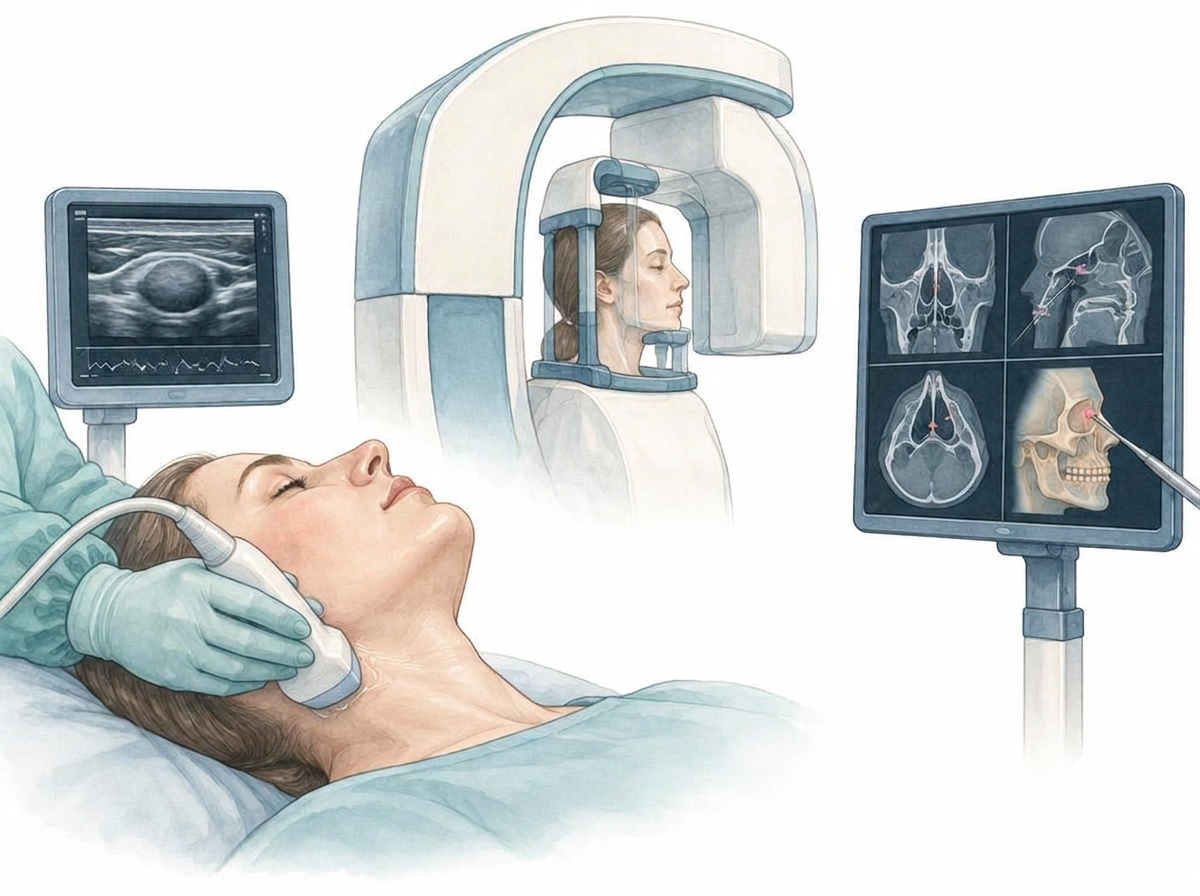
Ultrasound examinations (sonography) use sound waves and do not involve X-rays. In the head and neck they are used, among other things, to assess soft tissues such as the salivary glands, lymph nodes and changes in the neck. A clinician decides after examination whether an ultrasound examination is useful in your individual case. At the Kopfzentrum Bielefeld, examinations are performed with the ACUSON Maple ultrasound system from Siemens Healthineers.
What happens during the examination?
A gel is applied to the skin to improve sound contact. A small transducer is moved over the area being examined; the images appear in real time on a screen. The examination is painless and generally requires no special preparation. Duration depends on the clinical question.
Applications in the head and neck region
Typical questions involve soft tissues: swellings or changes in the salivary glands, enlarged or conspicuous lymph nodes in the neck, neck cysts and unclear lumps under the skin. Ultrasound can also be the first imaging method for swellings, for example in children. When a tissue sample is needed, fine-needle aspiration can be performed with targeted ultrasound guidance.
Limitations and complementary methods
Bone and air obstruct the transmission of sound, so structures behind them or in air-filled spaces cannot be fully assessed with ultrasound. For questions about bone, CT or cone-beam CT and MRI are available; for many soft-tissue questions MRI is particularly informative. The ultrasound findings are therefore always interpreted alongside your symptoms, the examination and other results; the team clarifies with you which method is appropriate in your case and where it takes place.
02
Radiology & cone-beam CT
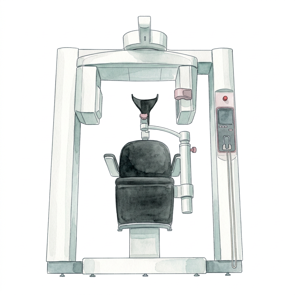
Cone-beam computed tomography (CBCT), known in German as digitale Volumentomographie (DVT), uses X-rays to create a three-dimensional view of a selected area of the head and face. It provides detailed information about bony structures in particular. A clinician decides whether CBCT or another imaging method is appropriate for the specific clinical question.
How are the three-dimensional images created?
An X-ray source and detector move around the head, capturing images from different angles. A computer reconstructs a volumetric dataset. It can be viewed in different cross-sectional planes and displayed as a three-dimensional model, allowing the position, shape and relationships of neighbouring structures to be assessed.
Applications in ENT and oral and maxillofacial care
Depending on the clinical question, CBCT can be used to assess the bony anatomy of the nose and sinuses, temporal bone, jaws and facial skeleton. Three-dimensional imaging may support the assessment of selected changes or injuries and the planning of a procedure. Not every symptom requires an X-ray examination; consultation and clinical examination come first.
Kopfzentrum describes the use of CT or CBCT image data for Fiagon surgical navigation in selected nose and sinus procedures. Instrument positions are related to the imaging dataset. Navigation supports orientation during surgery but does not replace clinical judgement.
Procedure and preparation
Before the scan, the clinical question and any previous imaging are reviewed. Tell the team if you are pregnant or might be pregnant. Jewellery, glasses or removable items in the scanned area may need to be removed to avoid interference with the images.
Your head is positioned for the area being examined. While the imaging unit moves, keep as still as possible and follow the team’s instructions. The scan itself is painless; contrast medium is generally not needed for a standard CBCT examination of bony structures. Exact positioning and duration depend on the scanner and imaging protocol.
Radiation protection and individual assessment
CBCT uses ionising X-rays and is therefore not radiation-free. Every examination needs a justified medical indication. The scan area and settings are selected to achieve the image quality needed for diagnosis with the lowest appropriate exposure. Careful assessment is particularly important in children and adolescents.
Radiation exposure depends on factors including the equipment, scan volume and settings. It is therefore not correct to claim that CBCT always involves less radiation than conventional CT. If pregnancy is possible, particular care is taken to assess whether the scan is necessary, can be postponed or can be replaced by a suitable alternative.
Limitations and alternative methods
CBCT is particularly useful for depicting bony structures. Other methods are more suitable for many questions involving soft tissues, nerves or tumour extent. Conventional CT may provide additional information depending on the clinical question; MRI does not use X-rays and is particularly helpful for many soft-tissue assessments. Ultrasound or endoscopy may also complement an examination or provide an alternative.
Results and next steps
The images are professionally interpreted and considered alongside your symptoms and other examination findings. This informs the next steps, such as further assessment, non-surgical treatment or surgical planning. Before your appointment, the team clarifies which examination is needed, where it will take place and any questions about cost coverage.
Frequently asked questions
This general information does not replace individual medical advice. Follow the instructions of your examining team.
When is instrumental diagnostics useful?
Ultrasound and CBCT answer different questions: ultrasound assesses soft tissue such as the salivary glands, lymph nodes or the thyroid, while CBCT shows bony structures and tooth positions. Neither replaces a medical examination – both supplement it. The physician selects the suitable method according to the findings and the clinical question.
Navigation in the operating theatre
For procedures close to critical structures, navigation in the operating theatre can display the position of the instruments in a three-dimensional image. The navigation systems at Kopfzentrum Bielefeld are described on the equipment page. To the equipment page