Home

Kopfzentrum Bielefeld

Equipment

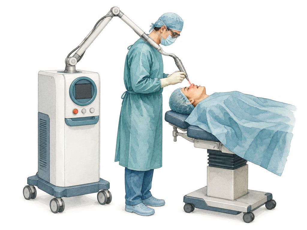
Equipment at Kopfzentrum Bielefeld: laser systems and navigation technology for selected ear, nose and throat procedures. The choice of system depends on the findings and the individual treatment plan.

Illustration: laser system with handpiece in the treatment room

Laser systems

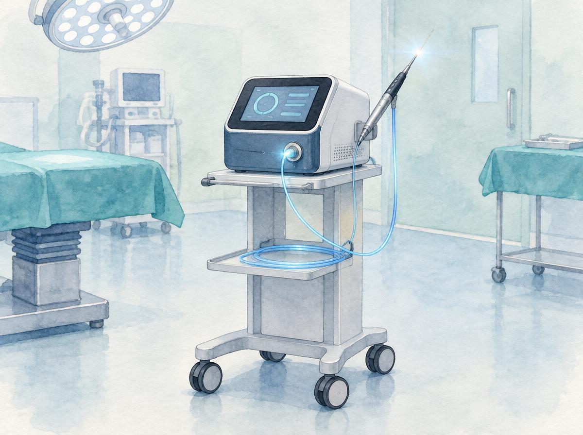
Colour illustration of a compact diode laser on a trolley in an operating room

LEONARDO diode laser with two wavelengths

The clinic uses the LEONARDO diode laser. This compact diode laser combines two wavelengths (980 nm and 1470 nm), which can be used individually or together.

The tissue effect can be matched to the intended goal – from precise cutting and vaporisation of tissue to haemostasis, including endoscopic procedures. Whether the method is suitable in an individual case is determined during medical treatment planning.
Further information and source
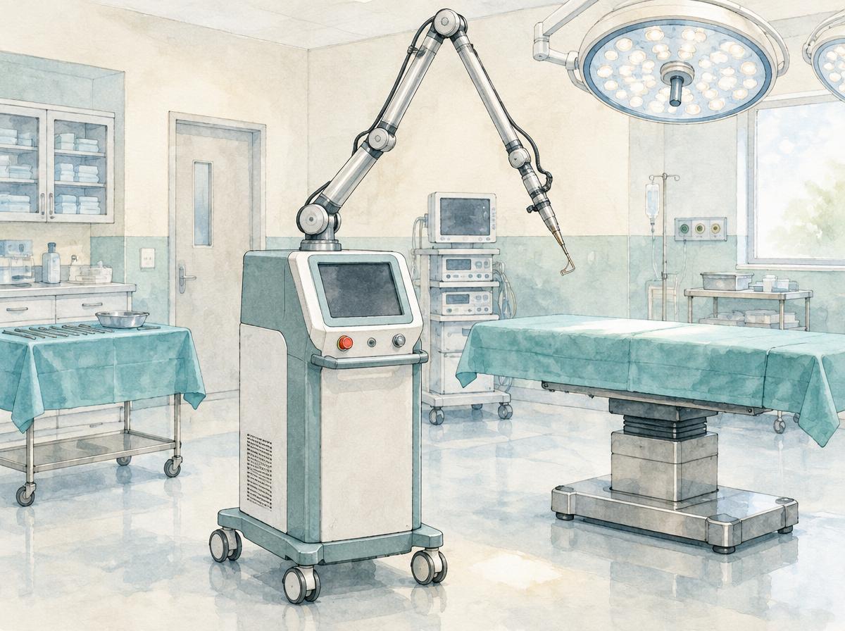
Colour illustration of a CO₂ laser with articulated arm in an operating room

Sharplan CO₂ laser for precise tissue incision

The Sharplan CO₂ laser operates at a wavelength of 10.6 µm and is used in ENT surgery, among other things, for precise incision of soft tissue.

The system offers continuous-wave and superpulse modes. The shallow penetration depth of the CO₂ laser allows fine incisions with little heat spread into the surrounding tissue. Whether the method is appropriate in an individual case is determined by medical treatment planning.

Further information and source
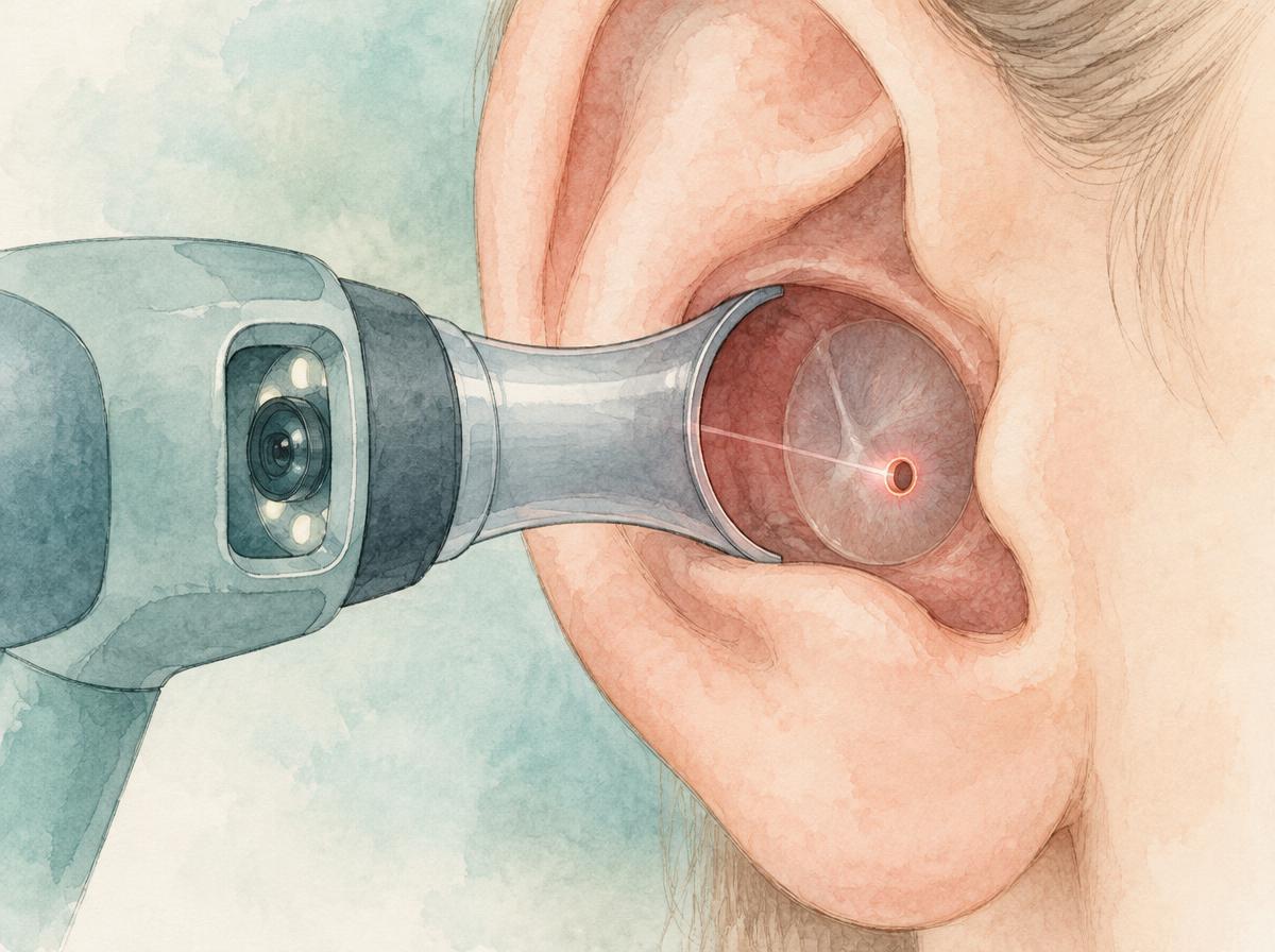
Colour illustration of a video otoscope at the ear canal with a small opening in the eardrum

Laser-assisted myringotomy with OtoScan

The OtoScan system combines a video otoscope with CO₂ laser delivery. It allows a pre-defined, precisely sized opening in the eardrum (myringotomy) without a scalpel; the diameter of the window can be set in small increments.

The procedure is considered, among other indications, for middle-ear effusions. Whether laser-assisted myringotomy is suitable in an individual case is determined during medical treatment planning.
Further information and source
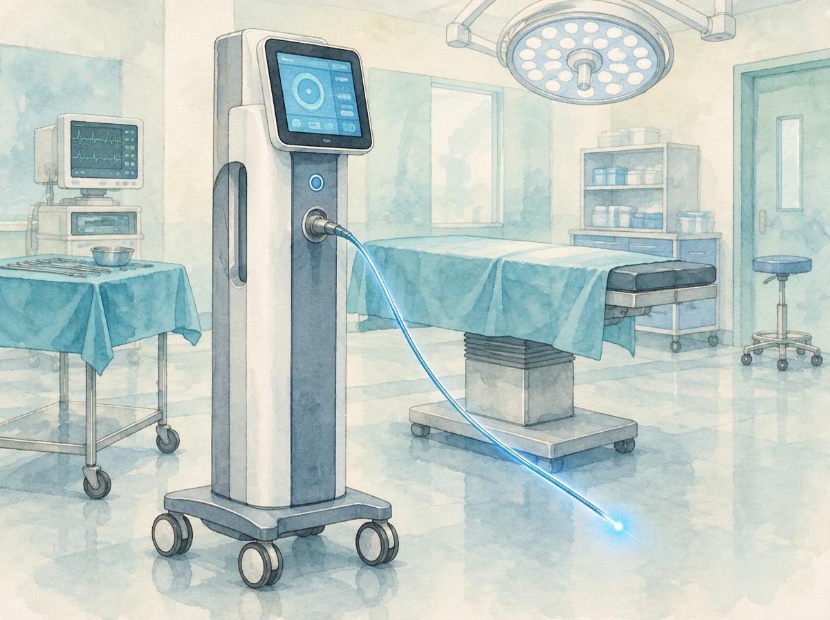
Colour illustration of a laser tower with a glowing laser fibre in an operating room

Laser technology by A.R.C. Laser – made in Germany

The clinic uses laser systems by A.R.C. Laser GmbH from Nuremberg. The portfolio covers a broad wavelength spectrum: from the classic C-Las CO₂ laser for extended procedures such as tumour resection, the FOX diode laser for middle-ear surgery, to the blue WOLF laser (445 nm) for phonosurgery and vascular tissue.

The devices are developed and manufactured in Germany. We introduced the TruBlue™ laser in a separate news item. Which variant is used in an individual case depends on the medical treatment plan.

Further information and source
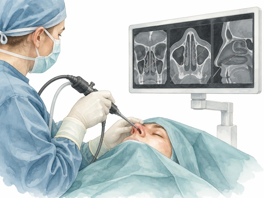
Illustration: navigated endoscope during a nasal procedure, monitor with three cross-sectional views of the sinuses

Fiagon XS navigation in nasal and sinus surgery

For selected operations of the nose and sinuses the clinic uses the Fiagon XS navigation system. Using CT or cone-beam CT images, it displays the position of the instruments in real time during the procedure.

In complex anatomy or after previous surgery this imaging can supplement orientation. Navigation does not replace the surgeon’s experience or clinical judgement.

Further information and source
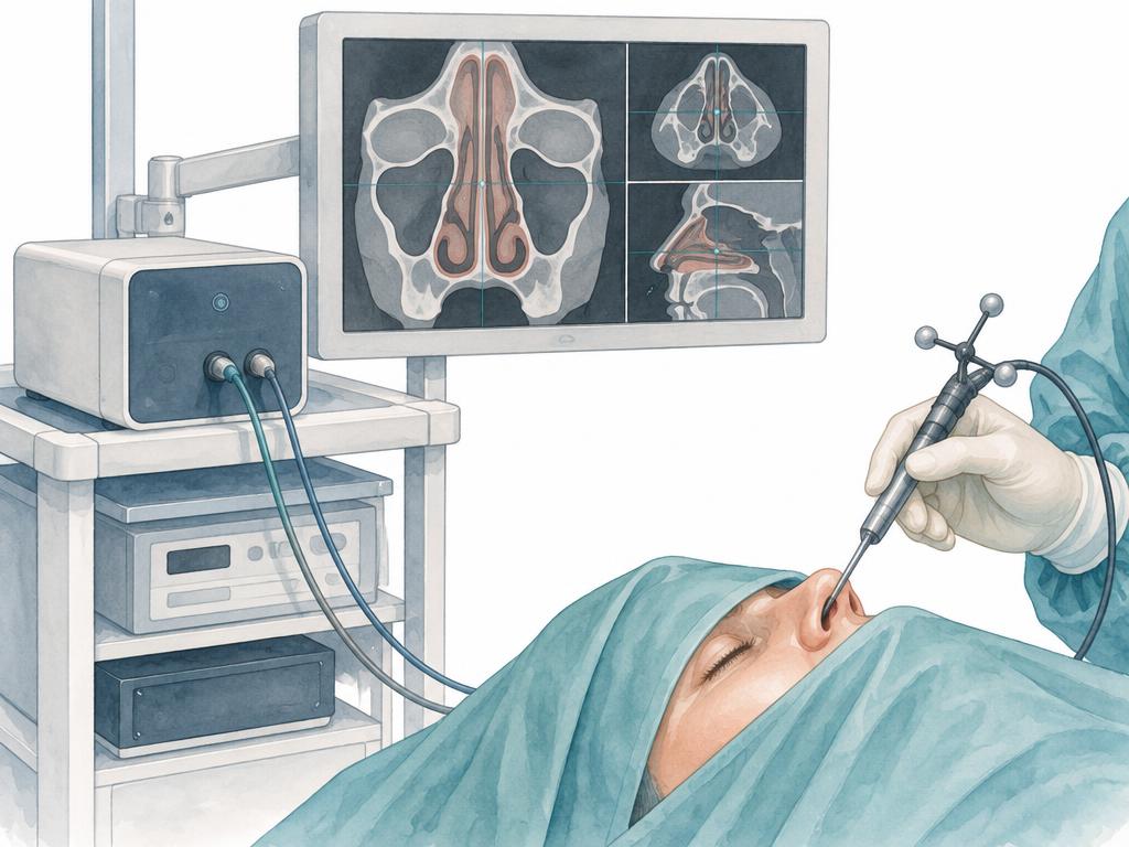
Illustration: compact navigation unit on an operating cart, a monitor with cross-sectional images beside it and a navigated instrument over a draped patient

Cube 4D navigation system

Cube 4D is Fiagon’s compact navigation unit. The system tracks instruments electromagnetically and can be fitted into existing operating carts and media towers.

Instrument positions are shown in relation to the patient’s own imaging, and the system is designed for procedures in the ENT field. Whether navigation is used in an individual case is decided during treatment planning.

Further information and source
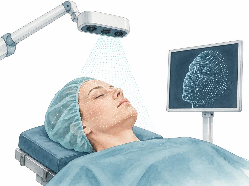
Illustration: camera on an arm above the face of a lying patient projecting a fine dot pattern onto the skin, with a monitor showing the three-dimensional face model

VirtuEye – contactless photo registration

VirtuEye belongs to the Fiagon navigation and establishes the link between patient and imaging. A camera captures the surface of the face with a large number of measurement points without touching the skin.

Registration takes place before the procedure begins; checking this match remains with the treatment team.

Further information and source

Neuromonitoring

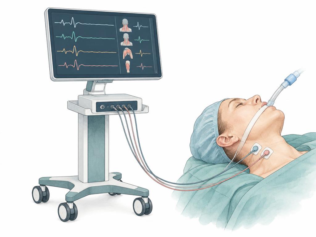
Illustration: neuromonitoring unit with a screen showing waveform curves, connected by cables to an intubated patient

Intraoperative neuromonitoring with AVALANCHE

During operations in which the function of a nerve matters for the outcome, that function can be measured while the procedure is under way. The clinic uses the AVALANCHE neuromonitoring system made by Dr. Langer Medical: fine electrodes record the muscle responses (EMG), the signals are displayed continuously, and a controlled stimulus can be used to demonstrate a nerve directly.

For surgery of the larynx and thyroid the vocal fold muscle is recorded through a special electrode tube, and during ear surgery the facial nerve can be controlled in the same way. Whether monitoring is used in an individual case, and which recordings are employed, is decided by the medical team during treatment planning.

Further information and source

Modern microscopes

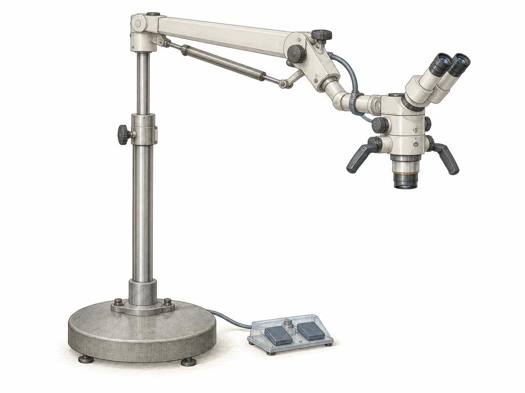
Illustration: surgical microscope on a tall floor stand with a multi-jointed boom arm, binocular tube and objective, a foot switch beside it

ZEISS S88 surgical microscope

Many procedures of the ear, skull base and neck can only be carried out safely when fine structures stay visible in magnification and reliably illuminated over a longer period. The clinic therefore uses a ZEISS surgical microscope mounted on the S88 floor stand. The optics work apochromatically, reproducing the surgical field with true colour; magnification is changed under motor drive, and focus is maintained by the internal focusing mechanism so that no objective lens has to be exchanged.

The heavy floor stand holds the microscope head steadily in the position once chosen by means of magnetically locking joints, yet allows it to be moved cleanly when required; a second tube can make the same view available to another person. Whether a surgical microscope is used in an individual case, and how it is set up, is decided by the medical team during treatment planning.

Further information and source
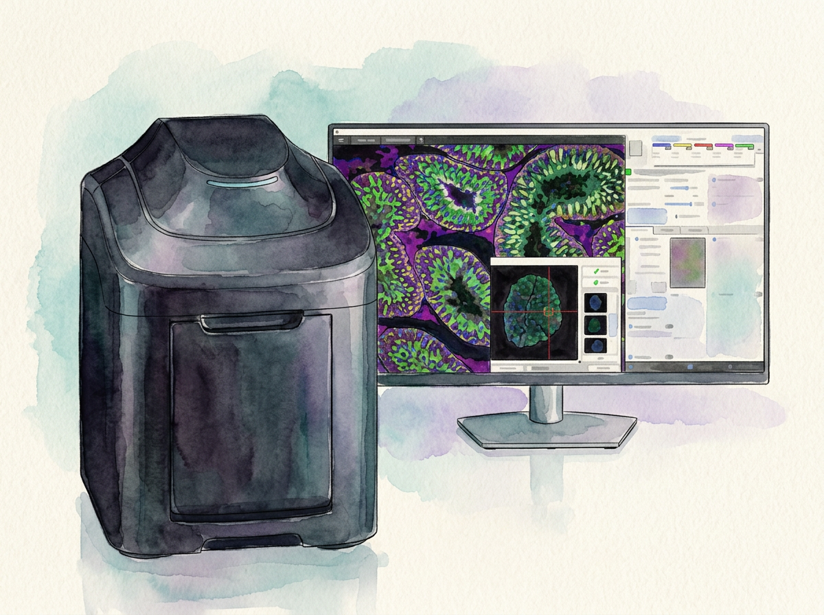
Illustration: Keyence research microscope with a monitor showing fluorescence scans

Keyence research microscope

For high-resolution imaging in its own laboratory the clinic uses a research microscope made by Keyence. The microscope is connected to its own monitor: the images are viewed, evaluated and archived on screen rather than assessed only through the eyepieces.

This includes fluorescence measurements across several channels. In this process labelled structures within a specimen are selectively excited and their signals recorded separately, so different features can be shown simultaneously in a single specimen and compared side by side on the monitor. Whether and in what form a microscopic examination takes place in an individual case is decided by the medical team.

Further information and source

4K video towers

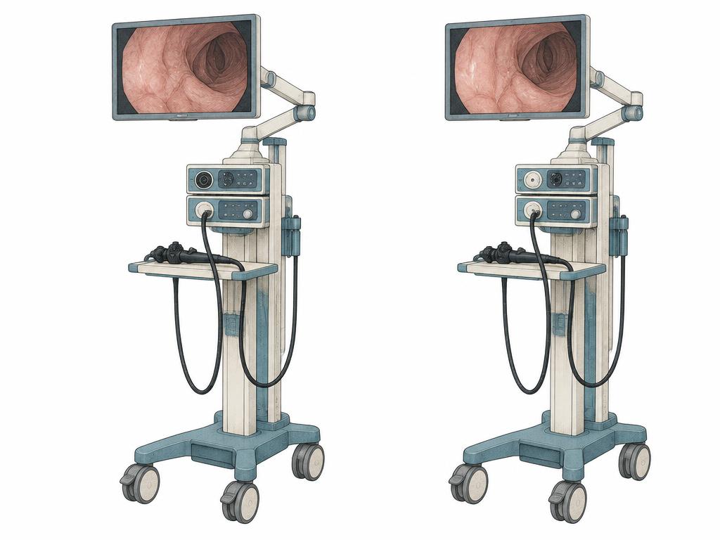
Illustration: two mobile video towers standing side by side with monitors on articulated arms, an endoscope on the tray and cables

Two Arthrex 4K video towers with Pano scope

Endoscopic procedures require a magnified and reliably illuminated view into spaces that cannot be seen with the naked eye. The clinic works with two Arthrex 4K video towers for this purpose. The systems combine camera, light source and image control in a single compact column; the images appear in high resolution on a large monitor that can be positioned freely within the surgeon’s field of view.

This includes the Pano scope: at the touch of a button it switches between a 30°, a 70° and an ultrawide view. More anatomy therefore becomes visible on the same monitor, and fewer additional portals are required. Having two units in the house allows the video technology to be used according to the procedure and the room. Whether such a system is used in an individual case, and with which settings, is decided by the medical team during treatment planning.

Further information and source

Ultrasonic surgery

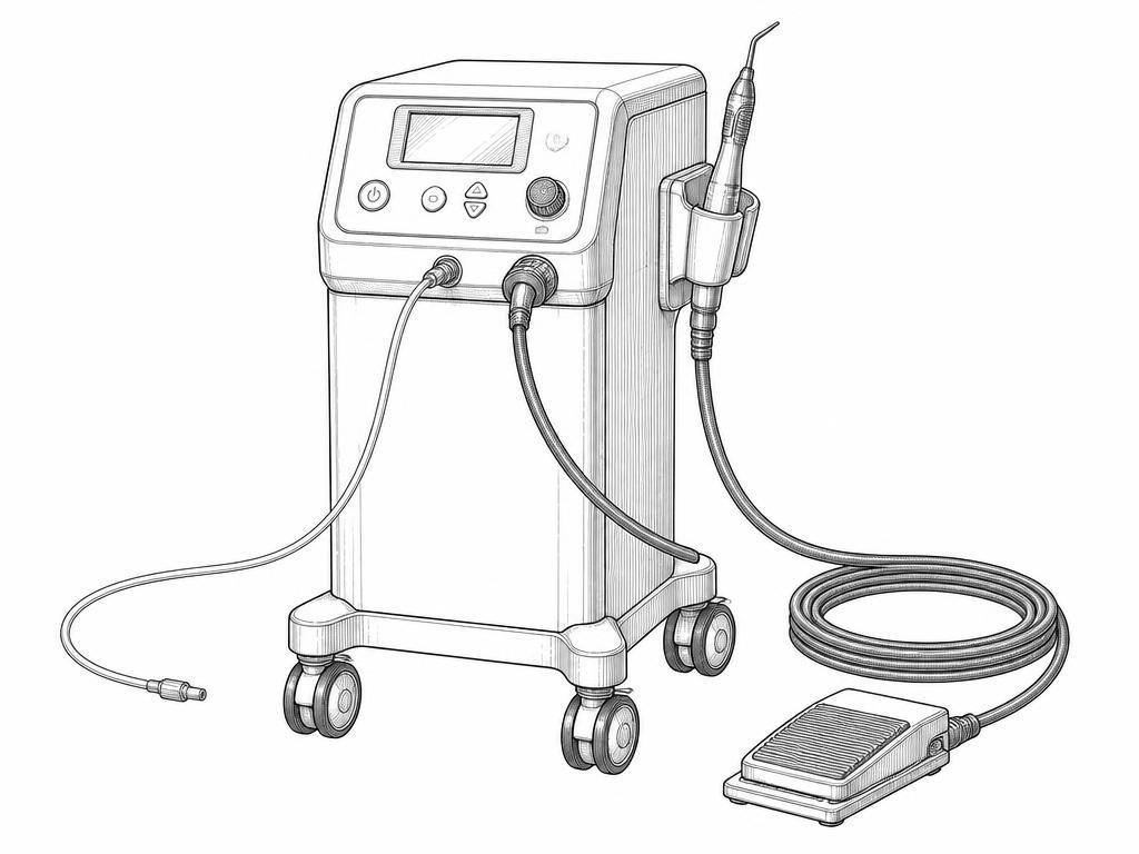
Illustration: mobile ultrasonic aspiration system with a control panel, a handpiece in its holder and a foot switch

Ultrasonic tissue removal with a Söring system

The clinic uses an ultrasonic aspiration system from Söring (Söring GmbH, Quickborn) for ultrasonic tissue removal. The fine tip of the handpiece delivers focused ultrasonic vibrations into the tissue, which selectively emulsify softer, water-rich components while firmer structures such as vessel walls and nerves are largely spared. The emulsified tissue is suctioned away at the same time and cooled through an irrigation.

The method is considered wherever delicate structures lie close by and tissue should be removed as gently as possible. Whether and in what form such a system is used in an individual case is decided by the medical team during treatment planning.

Further information and source