
Kopfzentrum Bielefeld
Tumour surgery
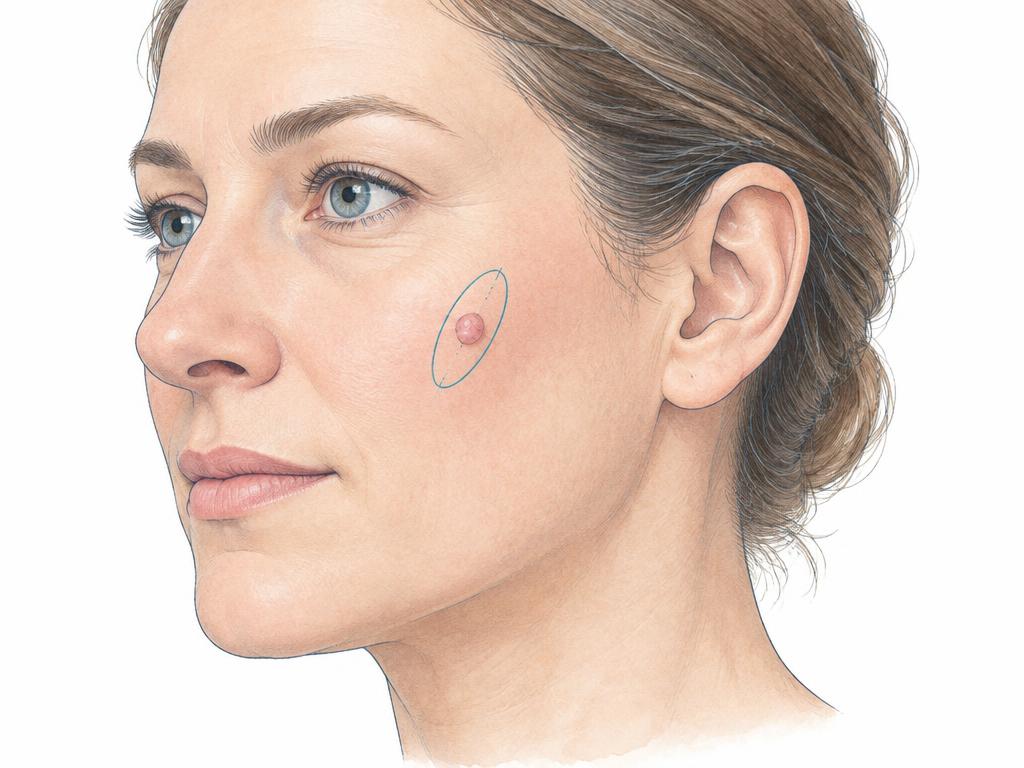
Diagnosis
Most skin changes are benign, but some are malignant and should therefore be removed early. To diagnose malignant tumours as early as possible, regular preventive examinations are important to identify any changes immediately and initiate the necessary treatment steps.
Prevention
If these skin tumours – particularly carcinomas and malignant melanoma – are detected and treated early, there is a very high chance of a complete cure. Skin cancer screening every two years from the age of 35 therefore offers a good preventive option.
Treatment options
Atheromas: Atheromas (also known as sebaceous cysts) arise from inflammation of the sebaceous glands. If the outlets of the sebaceous glands are blocked, secretions such as fats cannot drain outwards. This results in benign cysts (atheromas), mainly filled with fat, which can grow to the size of a hen’s egg.
Symptoms occur when the atheroma becomes inflamed or pus has accumulated in addition to the fat. Early removal is advisable, particularly to prevent unsightly scarring.
Basal cell carcinoma removal: Basal cell carcinomas often occur in fair-skinned people who expose themselves to intense and excessively frequent sunlight. They typically develop on areas of skin exposed to the sun (neck, head, hands).
Secondary tumours, known as metastases, generally do not occur with basal cell carcinomas. The affected areas of skin are cut out generously with a scalpel: this is the best method for removing the tumorous skin lesion in the long term.
The removed areas of skin are sent to the laboratory for additional examination by a tissue specialist (pathologist). This allows us to establish with certainty which tumour was present and whether we were able to remove it completely.
Carcinoma removal: Carcinomas (or squamous cell carcinomas) are mostly found in older people living in hot, sunny regions. Like basal cell carcinomas, they often occur on areas of skin frequently exposed to sunlight (neck, head, backs of the hands).
Secondary tumours, or metastases, are comparatively rare in carcinomas. The affected areas of skin are removed with a scalpel to enable a lasting cure. We send the tissue samples to the laboratory for examination by a pathologist, who can check whether we have completely excised the carcinoma.
Removal of malignant melanoma: Malignant melanoma (also called black skin cancer because of its colour) is the most malignant skin tumour. However, if it is detected sufficiently early, the prognosis for a cure is generally very good.
In particular, people who frequently had sunburn during childhood and adolescence, have fair skin, live in sunny regions or have family members who have had malignant melanoma should examine their skin for changes themselves approximately every four weeks.
The entire malignant melanoma is removed in a surgical procedure. Regular follow-up appointments are particularly essential here.
Oral cavity tumours (tumours of the floor of the mouth, upper jaw and lower jaw): Malignant tumours can also occur in the floor of the mouth, on the tongue or in the upper and lower jaws. Initial signs may include sore areas in the mouth that have not healed after around one to two weeks (and are mistaken for pressure sores caused by dentures). This is why preventive examinations at your dentist are so important: the dentist is often the first to notice that a change has developed.
If a tumour is present, it is important to remove it completely in a surgical procedure, along with any extensions and secondary tumours. In the neck, for example, lymph vessels and lymph nodes are often removed as well, as secondary tumours frequently spread via these structures (this is known as “neck dissection” or submandibular clearance). During these operations, care is taken to remove the tumours completely while preserving the shape and function of the facial areas.
If this was not possible, they are subsequently reconstructed using plastic surgery techniques. For areas of jawbone, for example, bone can be transplanted from the pelvis or ribs. For areas of skin, tissue can be transplanted from the forearm, back or chest.
Depending on the findings, radiotherapy or chemotherapy may be necessary afterwards.
After the procedure
After treatment, continue to have regular examinations by your dermatologist for possible pathological skin changes. Statistically, people who have already been affected have a higher likelihood of developing another skin lesion.
Malignant melanoma in particular can form metastases. It is therefore essential to arrange regular follow-up appointments with your treating dermatologist.
Other information
Tips for early detection: Have you recently noticed that a mole or another mark on your skin has changed in colour, shape and size? This could be a sign that a previously benign skin lesion has become malignant.
Other possible signs include increased crusting, hardening, a tendency to bleed or weeping. Have your dermatologist or GP check whether a malignant skin lesion is responsible.
The so-called ABCDE rule can also give you initial indications:
Asymmetry: Is the skin lesion asymmetrical, rather than circular? It could then develop into a malignant skin lesion. Symmetrical, meaning circular, lesions, on the other hand, are usually benign.
Border: Jagged, irregular or blurred edges of a “mole” can indicate a pathological development. If the skin lesion is sharply demarcated from your normal skin, it is usually benign.
Colour: Skin lesions with several colours (brown, black, grey, white, red or blue) frequently indicate a malignant tumour. If the colour remains the same over a longer period (usually black or brown), this is usually no cause for concern.
Diameter: Rule of thumb: All moles under five millimetres in diameter are normally benign. Those that grow larger than five millimetres or are already larger may be malignant and should be checked.
Elevation: If a skin lesion has developed within a few weeks on previously smooth skin, or if the skin lesion is growing upwards, this may indicate disease.