
Kopfzentrum Bielefeld
Orbital floor fracture
Diagnosis
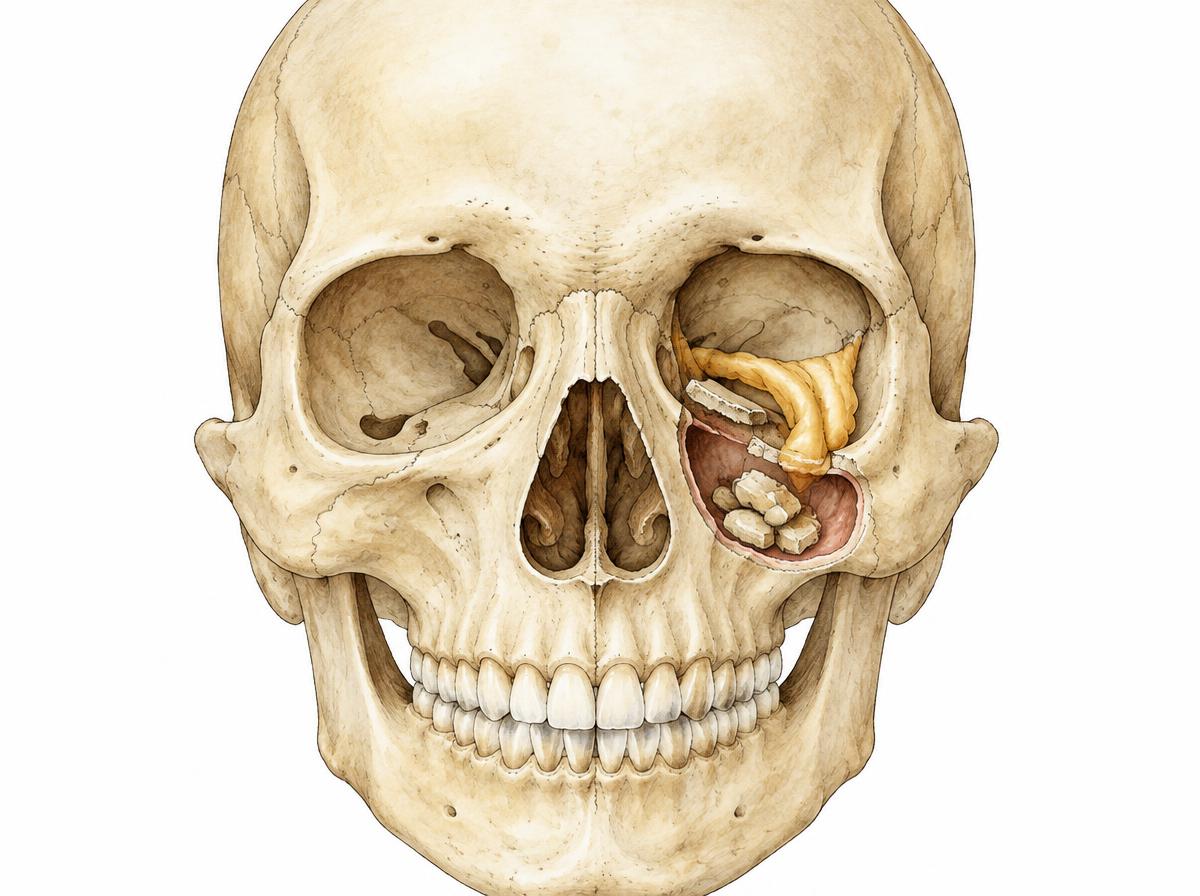
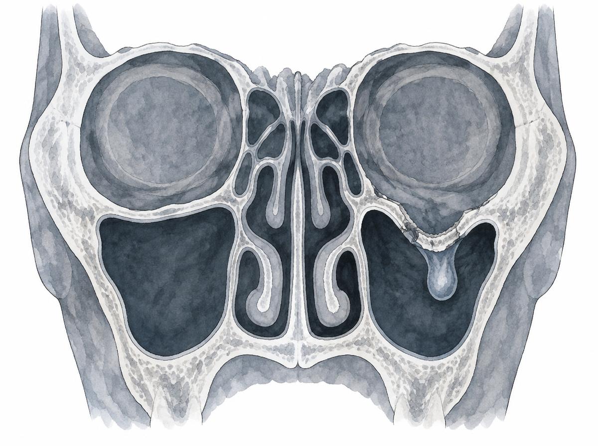
In an orbital floor fracture, the thin bone beneath the eye breaks, usually from a blow or fall onto the eye and cheek. Possible symptoms include swelling and bruising, double vision when looking up or down, numbness of the cheek and upper lip, and a sunken eyeball. Diagnosis is made by clinical examination, assessment of eye movement and cross-sectional imaging such as CT or cone-beam CT.
Treatment options
Small fractures without double vision or a sunken eyeball can often be managed conservatively with follow-up. If eye muscles or tissue are trapped or the defect is larger, the orbital floor is reconstructed surgically, for example through an inconspicuous incision on the inside of the lower eyelid, and supported with a plate or implant. Whether and when surgery is advisable is decided individually and, if required, together with ophthalmology.