
Kopfzentrum Bielefeld
Diagnostics
Examination, surgery and inpatient treatment all take place at Kopfzentrum Bielefeld. The methods used depend on your symptoms and the findings of the medical examination.
01
Flexible digital video endoscopy
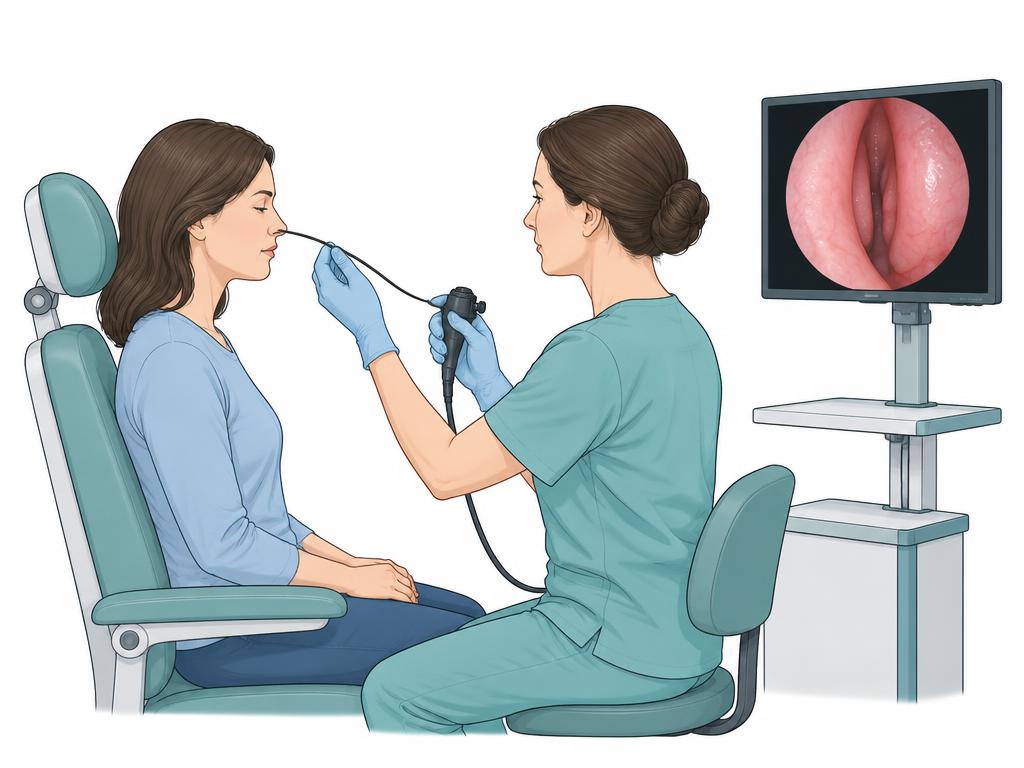
A very thin, flexible endoscope with a tiny camera allows the nasal cavities, nasopharynx, throat and larynx to be viewed from the inside. The image appears on a monitor, so the findings and the examination can be looked at together. The test usually takes only a few minutes.
Procedure
Before the examination the nasal lining is briefly sprayed: the spray makes the mucosa swell down slightly and numbs the surface. The flexible endoscope is then gently passed along the floor of the nose. This allows the septum, the turbinates, the narrow passages to the sinus openings and the nasopharynx to be assessed. The throat and larynx can be viewed afterwards.
What the examination shows
Endoscopy reveals areas that cannot be seen from outside: swollen mucosa, narrowings, polyps, mucus tracks, changes at the Eustachian tube opening and findings at the larynx. If needed, cell material can be collected during the examination.
In chronic rhinosinusitis, a clear endoscopic finding can shorten further work-up: according to a specialist review, computed tomography is not necessarily required when nasal endoscopy is positive. Whether imaging is needed is decided by the physician based on findings and course.
Larynx and voice
In persistent hoarseness, viewing the larynx is part of the work-up: the international guideline on dysphonia recommends visualising the larynx when hoarseness lasts longer than four weeks. Mobility, position and surface of the vocal folds can be assessed endoscopically; additional methods exist for the fine movement of the vocal folds.
Frequently asked questions
This general information does not replace individual medical advice. Follow the instructions of your examining team.
02
Hearing assessment
Behavioural hearing tests and objective measurements complement each other. Selection depends on age, symptoms and examination findings; no single measurement provides a complete hearing diagnosis.
Pure-tone audiometry
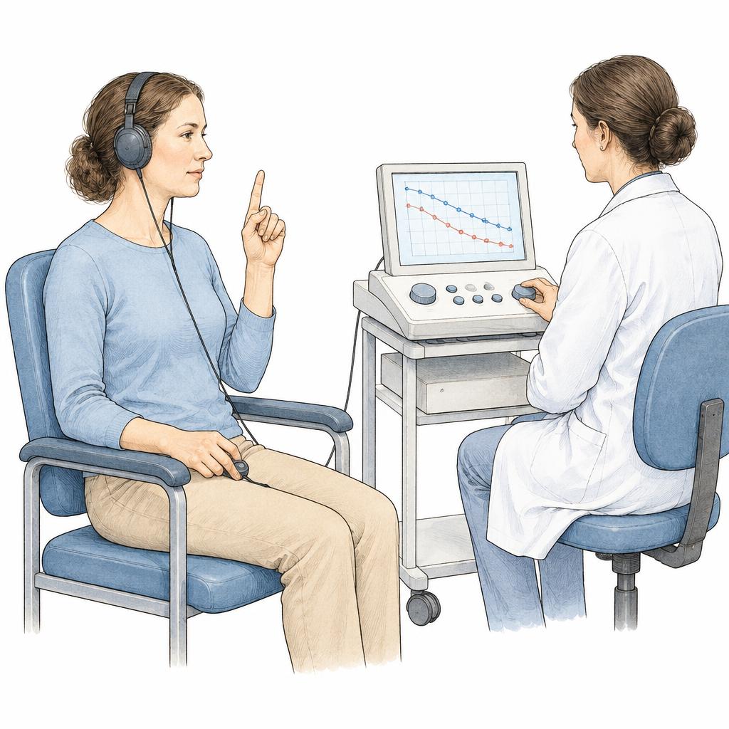
Headphones present tones at different pitches and volumes. You indicate when you hear a tone. The results form an audiogram showing hearing thresholds. Bone-conduction testing may be added when needed.
Speech audiometry
You listen to words or numbers and repeat what you understood. The test measures speech recognition at different sound levels; testing in background noise may be added depending on the clinical question.
Tympanometry
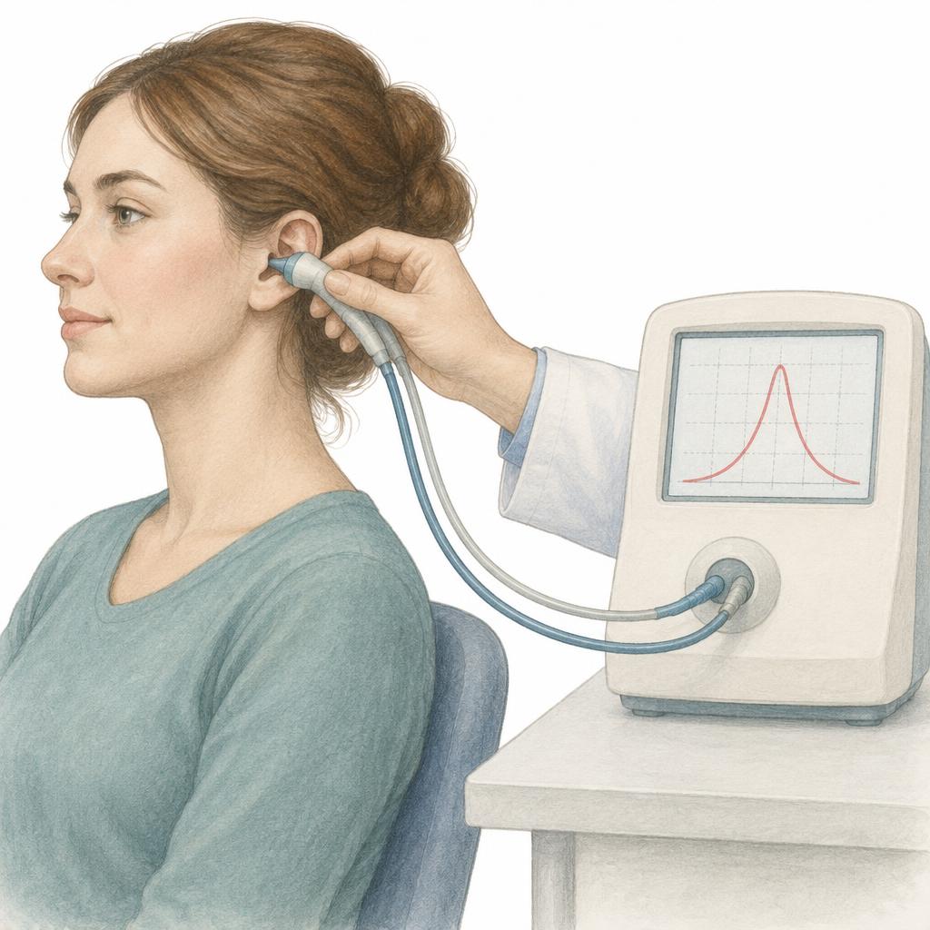
A probe in the ear canal briefly changes air pressure and measures eardrum and middle-ear mobility. It can indicate middle-ear fluid or ventilation problems, but does not directly measure hearing ability.
OAE – otoacoustic emissions
A small probe records faint sound responses from the cochlea after a sound stimulus. This objective measurement provides information about outer hair cell function. Middle-ear problems can affect the result; OAEs alone do not rule out every hearing disorder.
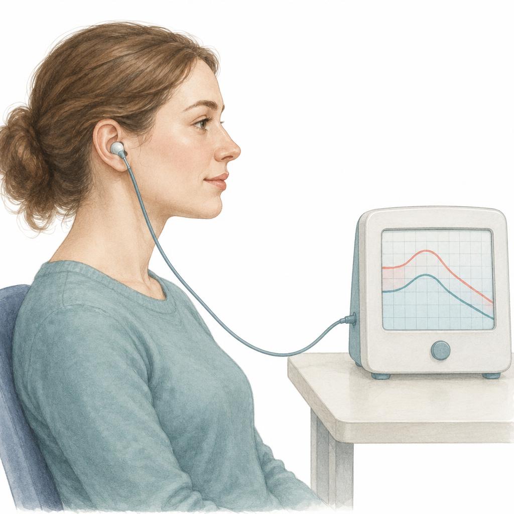
DPOAE – distortion-product OAEs
Two tones are presented together. The probe measures additional sound responses generated in the cochlea. DPOAEs allow frequency-specific assessment of outer hair cell function.
TOAE / TEOAE – transient-evoked OAEs
Brief sounds, usually clicks, produce measurable cochlear responses. The standard abbreviation is TEOAE. The method is used in hearing screening, among other applications, and complements other hearing tests.
Threshold BERA / auditory brainstem response (ABR)
Skin electrodes record electrical responses along the auditory pathway to the brainstem after sound stimulation. Varying the sound level allows hearing thresholds to be estimated. The test requires quiet rest and is particularly helpful when reliable behavioural responses cannot be obtained.
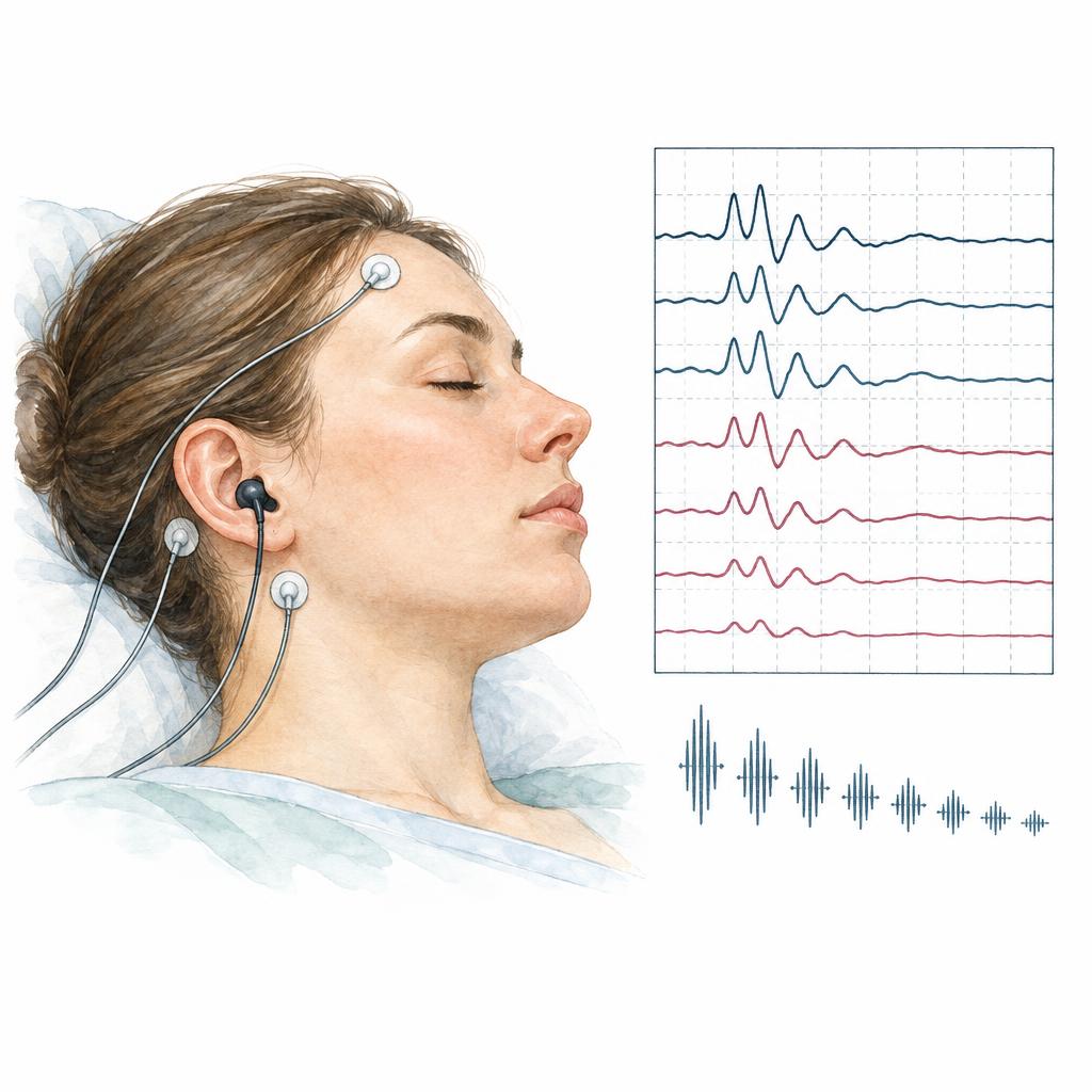
Electrocochleography (ECoG)
Electrodes record sound-evoked electrical potentials from the cochlea and adjacent auditory nerve. Electrode placement depends on the technique and is explained beforehand. The measurement can support selected inner-ear assessments, but does not by itself establish a diagnosis of Ménière’s disease.
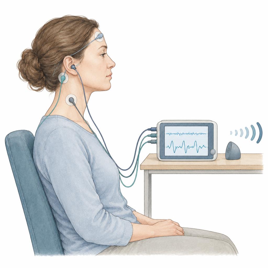
Tinnitus assessment
Tinnitus is the perception of a sound without an external source. Determining it includes an accurate description of the sound and measurement of its pitch and loudness: in tinnitus matching, a comparison tone is adjusted until it matches the sound you perceive. The level at which a noise covers the tinnitus fully or partly can also be recorded.
Because tinnitus frequently occurs together with hearing loss, pure-tone and speech audiometry form part of the assessment. Standardised questionnaires record how far the tinnitus affects daily life and make changes over time comparable. These measurements describe the tinnitus but do not by themselves reveal its cause. A newly developed, one-sided or pulsating tinnitus, and tinnitus with sudden hearing loss, should be assessed by a clinician promptly.
Medical information and sources
- ASHA – pure-tone and speech testing ↗
- UCLH – hearing and balance tests ↗
- Children’s Health – OAE, DPOAE and TEOAE ↗
- ASHA – auditory brainstem response ↗
- Weill Cornell – electrocochleography ↗
- Kopfzentrum Bielefeld – hearing disorders and tinnitus assessment (German) ↗
- European tinnitus guideline – diagnostics and assessment ↗
- NICE – tinnitus: assessment and management ↗
03
Dizziness and balance
Balance involves the inner ear, vision and body-position sensation. The following methods examine different parts of this system and are interpreted alongside medical history and clinical examination.
Unterberger / Fukuda stepping test
With the examination team ensuring your safety, you step in place with your eyes closed. Rotation or deviation is observed. This is a screening adjunct and cannot reliably localise a balance disorder on its own.
Romberg test
The team observes your stance with your feet close together, first with your eyes open and then closed. Changes in stability provide information about how sensory systems work together. The test is not specific to inner-ear disease.
cVEMP – cervical vestibular evoked myogenic potentials
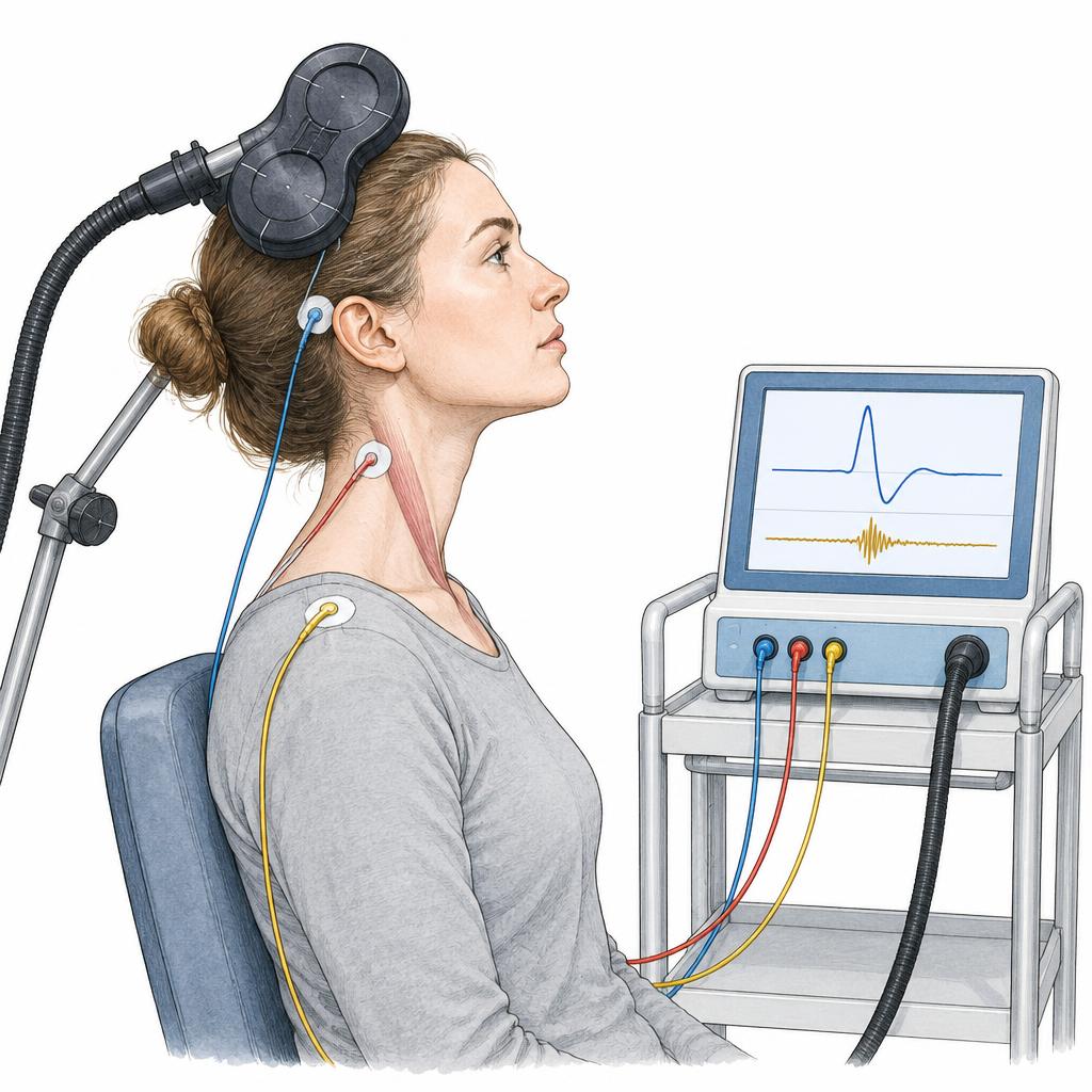
Sound or vibration stimuli evoke reflex responses recorded by skin electrodes over a contracted neck muscle. cVEMPs primarily assess the saccule and associated vestibular reflex pathways.
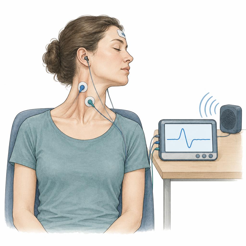
oVEMP – ocular vestibular evoked myogenic potentials
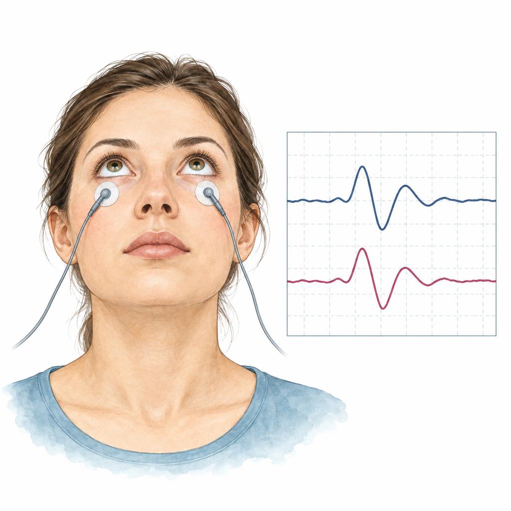
Electrodes below the eyes record reflex responses to sound or vibration while you look upwards. oVEMPs primarily provide information about the utricle and associated reflex pathways. Both VEMP methods complement other balance examinations.
Videonystagmography (VNG)
In videonystagmography, goggles with built-in cameras record your eye movements. The examination has several parts: following target lights, testing rapid eye movements (saccades), positional testing in different head positions and – if the clinician requests it – caloric testing, in which warm or cool airflow briefly triggers a spinning sensation. The direction, shape and strength of these eye movements indicate whether the balance organ on one or both sides responds abnormally.
The VNG is painless and usually takes one to two hours. Before the test, avoid make-up, alcohol and caffeine. Certain medications can affect the result; ask your clinician beforehand whether any should be paused, and do not stop anything on your own. Brief light-headedness or nausea can occur, so arranging a ride home is advisable.
Ocular counter-roll test
In the ocular counter-roll test, video goggles record how the eyes rotate when the head or body is tilted sideways: they turn slightly in the opposite direction as a reflex. The measurement provides information about the otolith-mediated perception of gravity and body orientation, mainly of the utricle. It complements other balance examinations, does not replace imaging and is not needed for every form of dizziness.
Transcranial magnetic stimulation (TMS)
In transcranial magnetic stimulation, a coil held over the head delivers brief magnetic pulses that activate the cerebral cortex, and electrodes record the resulting muscle responses. In ENT care the method has so far been used mainly in research, to study connections between the balance organs and the brain. It is not part of routine diagnostics in an ENT clinic and is relevant chiefly for neurological or scientific questions.
Contraindications must be clarified before use, for example epilepsy, a brain stimulator or metal implants in the head area. Whether such an examination is needed at all is decided by a clinician after clinical assessment; it is carried out in neurology or research departments equipped for it.
Head impulse test
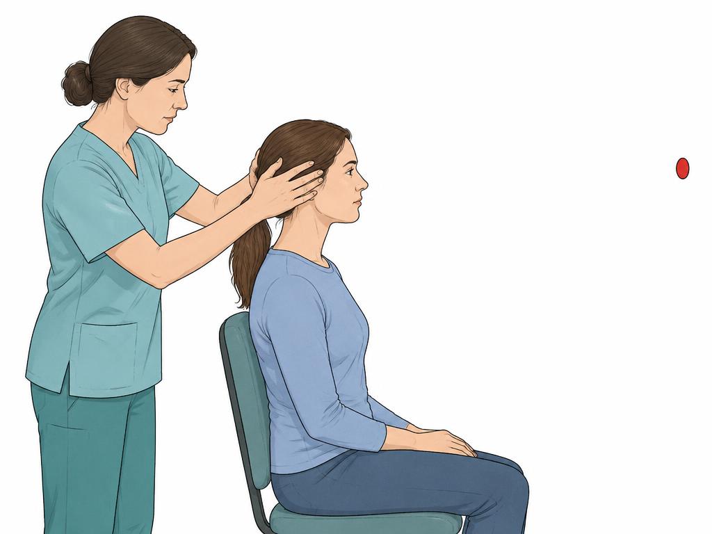
For the head impulse test you keep your gaze on a fixed point while the clinician quickly and gently turns your head to the left or the right. If balance function is intact, the eyes follow this movement automatically without losing the target. If the movement is not compensated, the eye makes a small corrective movement – an indication that the balance organ on that side may be working less well.
In the video-assisted form (vHIT), the eye movements are recorded with small goggles. This allows the gaze-following response to be measured for each individual semicircular canal, and even small corrective movements are detected. The test takes only a few minutes and complements other examinations such as caloric testing – it does not replace them.
Medical information and sources
- Stanford Medicine – vestibular testing ↗
- ASHA – dizziness and balance ↗
- MedlinePlus – videonystagmography ↗
- Brigham and Women’s – VNG test preparation ↗
- Research publication – video ocular counter roll as a test of otolith function ↗
- Scoping review – transcranial magnetic stimulation in chronic vestibular disorders ↗
- Review article – video head impulse test (English) ↗
04
Nose, nasal breathing and tubomanometry
The ear canal and eardrum are examined microscopically, and the nose and nasopharynx endoscopically. Additional functional measurements may be useful when symptoms suggest a Eustachian tube ventilation problem. A clinician decides after examination whether a measurement of nasal breathing is needed for your specific question.
Tubomanometry
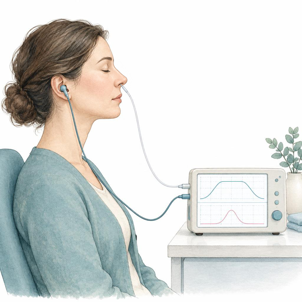
While you swallow, a controlled air pressure is applied through the nose. A measurement at the ear assesses whether and when the Eustachian tube opens and transmits pressure to the middle ear. Results are considered alongside symptoms, eardrum findings and other tests; this measurement alone does not prove or exclude Eustachian tube dysfunction.
Rhinomanometry
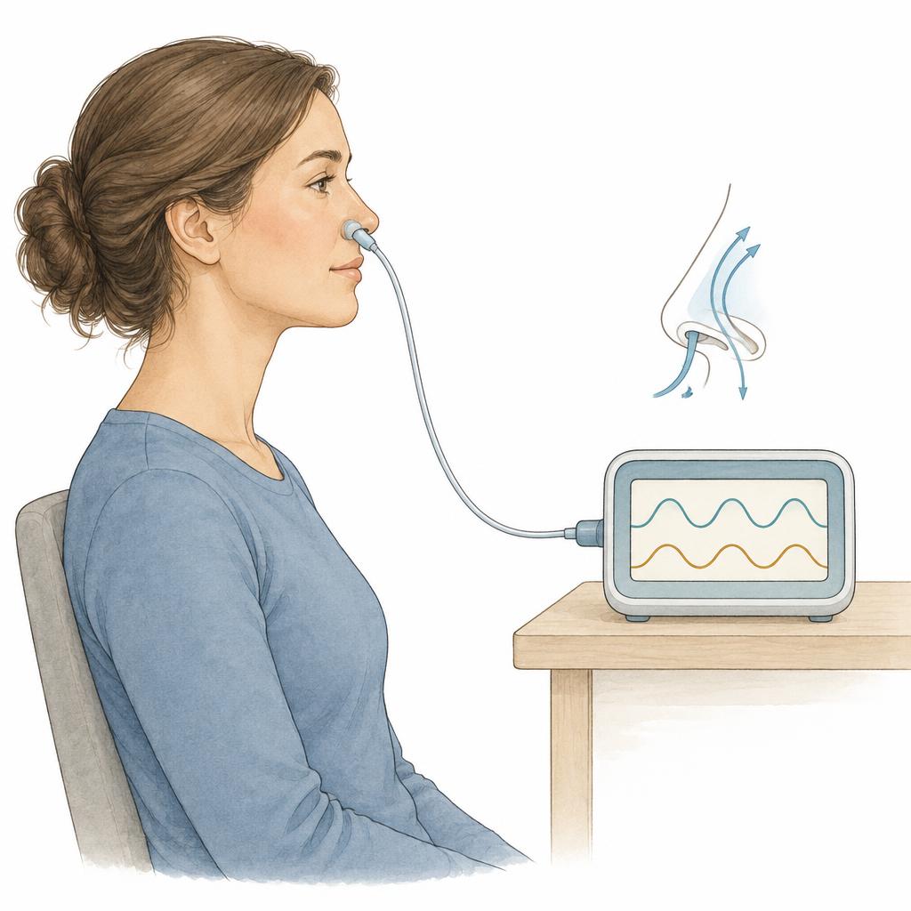
Active anterior rhinomanometry measures air pressure in front of the nostril and the airflow while you breathe normally. Both values are used to calculate the resistance of nasal breathing, separately for the right and left side. The figure shows how easy or how laboured breathing is at that moment. Results depend on how swollen the nasal lining currently is and can change over the day; they are interpreted alongside your symptoms and the endoscopic findings.
Rhinoresistometry
Rhinoresistometry records nasal resistance continuously across the breathing cycle rather than as a single figure. This shows how resistance changes during inhalation and exhalation. The display can indicate an unstable nasal airway that narrows while you breathe. It too cannot by itself establish where a narrowing lies, and is interpreted together with endoscopy and rhinomanometry.
Acoustic rhinometry
Acoustic rhinometry – sometimes called acoustic rhinomanometry – works with sound pulses: a brief tone is sent into the nose and a device calculates, from the returning echoes, how large the cross-section of the nasal cavity is at different distances. This provides the narrowest cross-sectional area and the volume of the nasal cavity. The method describes the shape of the nasal airway, but not airflow or resistance, and is therefore often combined with rhinomanometry.
FeNO measurement
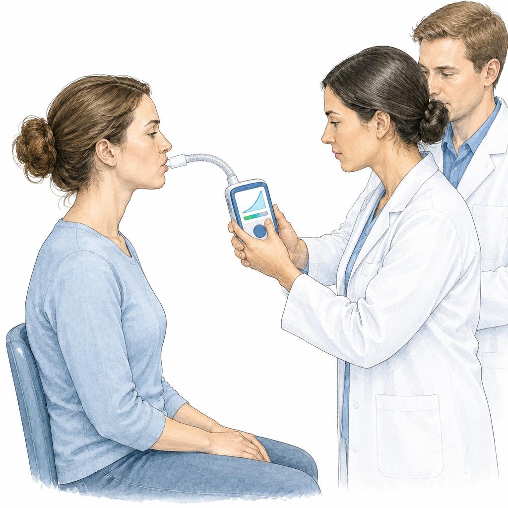
For the FeNO measurement you breathe calmly and steadily into a small device, which analyses the proportion of nitric oxide (NO) in your exhaled breath. The test is non-invasive, takes only a few minutes and complements lung function testing – it does not replace it.
An elevated value can point to a particular type of airway inflammation, as seen for example in allergic asthma. In adults a value below 25 ppb is considered low and a value above 50 ppb high. The FeNO value alone does not establish a diagnosis; the medical team interprets it together with the examination, lung function and other findings.
Medical information and sources
- Kopfzentrum – Eustachian tube dysfunction (German) ↗
- Research publication – tubomanometry ↗
- Review – methods for objectively assessing nasal patency (German) ↗
- Consensus report – rhinomanometry and acoustic rhinometry ↗
- RIGA consensus – nasal airway function tests ↗
- Clinical practice guideline – interpretation of exhaled nitric oxide levels (ATS) ↗
05
Allergies
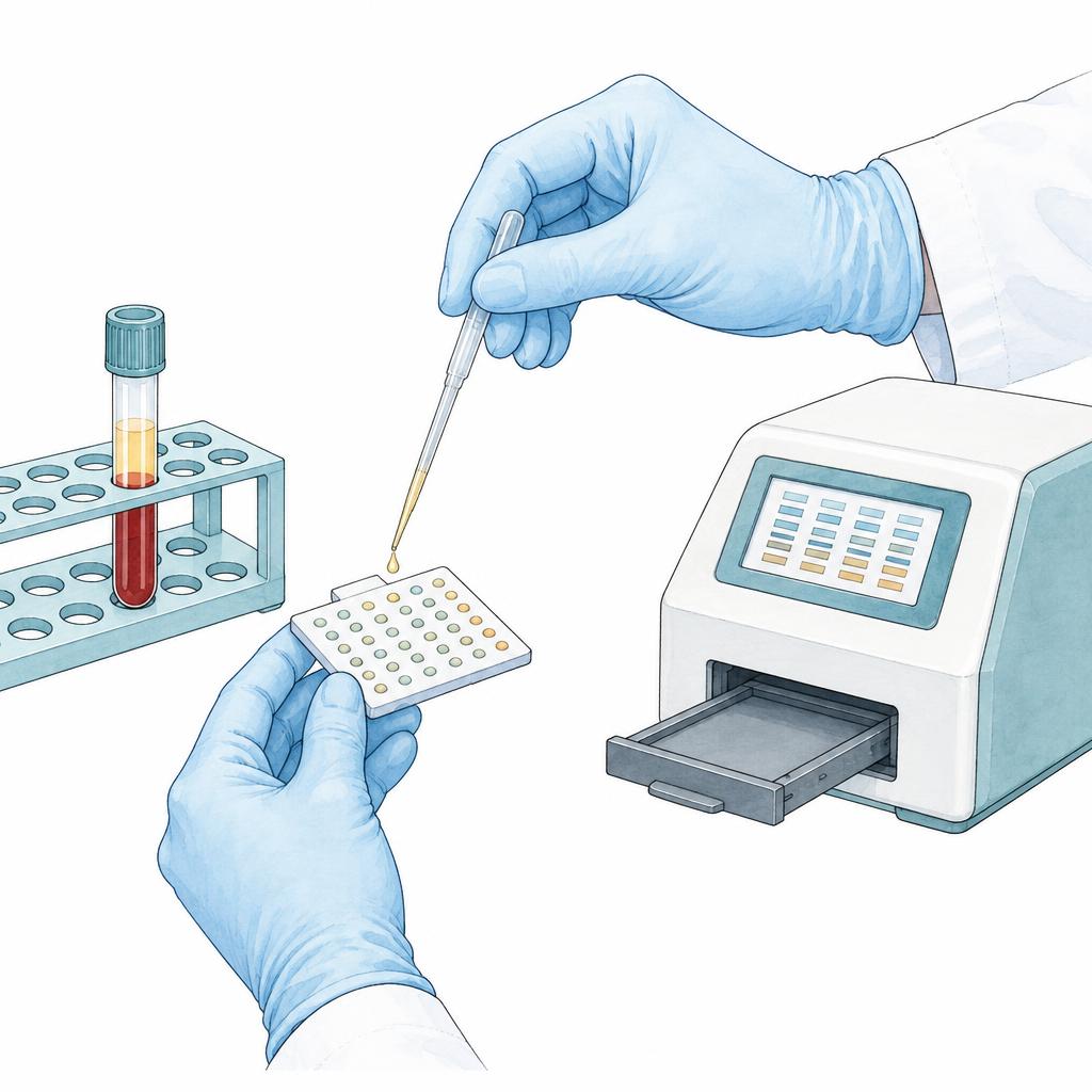
The MADx allergy test can examine antibodies against 295 allergens from a single blood sample. The results help to classify possible triggers and to plan further treatment.
06
Sleep
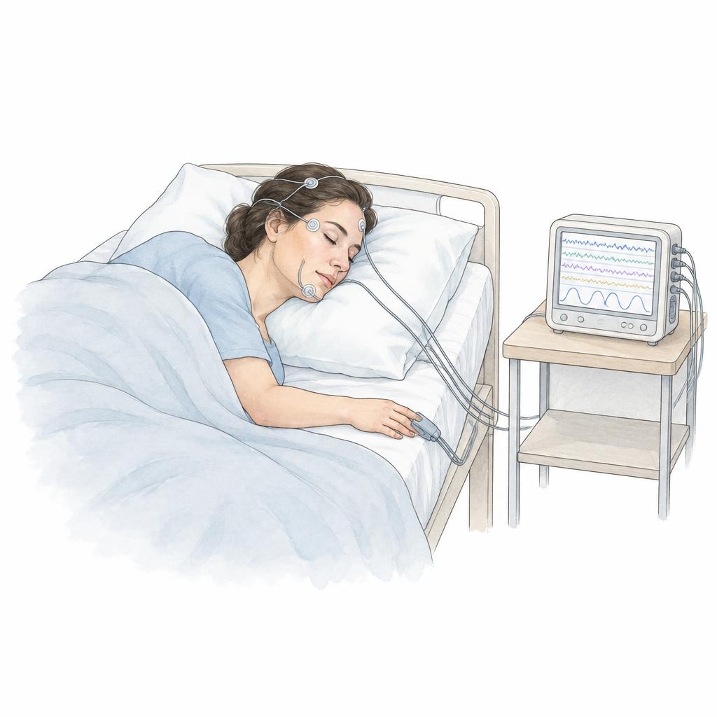
For the assessment of snoring and sleep apnoea, polysomnography is available – with a home device for sleep monitoring at home, or as complete inpatient sleep monitoring. Drug-induced sleep endoscopy (DISE) allows the behaviour of the airways during sleep to be assessed.
07
Skin
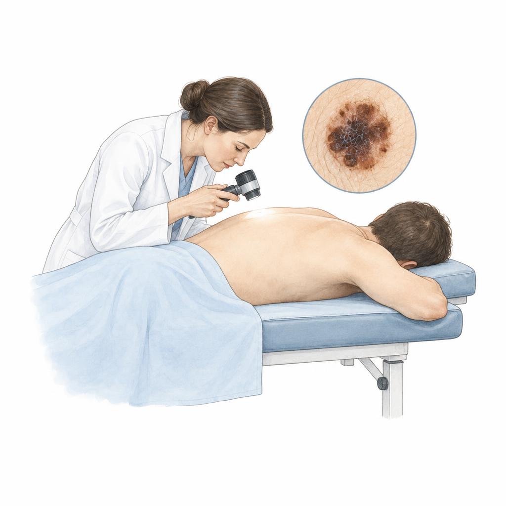
In skin cancer screening, skin changes are photographed in high resolution and compared with previous images using software support. In addition, we use an AI-supported analysis that assists the medical assessment; the final judgement is made by the specialist.
08
Head and neck
For the assessment of changes in the head and neck region, endoscopy including panendoscopy, fine-needle aspiration or tissue sampling and blood tests can be used. Depending on the findings, additional imaging such as CT, MRI or PET is arranged.
09
Hyperbaric chamber – treatment
Hyperbaric oxygen therapy is not a diagnostic examination. Medical oxygen is breathed in a chamber at increased ambient pressure. Whether this adjunctive treatment is appropriate requires a separate individual medical assessment.
Indication, preparation and risks
Before treatment, the specific indication, possible contraindications and ability to equalise pressure are assessed. Risks include pressure injuries to the ears and sinuses; other adverse effects are also possible. Benefits, alternatives, the procedure and cost coverage are discussed in advance. The team clarifies whether and where chamber treatment can be arranged.
Medical information and sources
10
Smell and taste
A declining sense of smell also dulls the perception of flavour and weakens the nose’s warning function — for example with smoke or spoiled food. Testing uses standardised test sets, and results are interpreted alongside nasal endoscopy and medical history.
Smell testing (olfactory assessment)
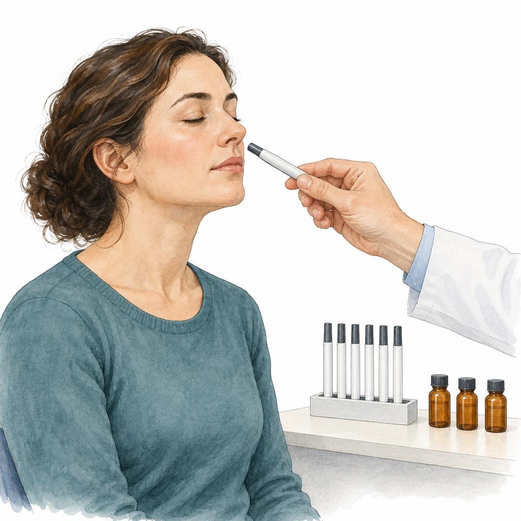
Three aspects are tested: recognising odours (identification), telling similar odours apart (discrimination) and the lowest concentration that can still be detected (threshold). Standardised test sets – for example scent pens or scent cards – provide comparable results that are checked against age references. The test requires reasonably free nasal breathing; during a cold or immediately after a decongestant nasal spray it is repeated at another time.
Taste testing
For the basic qualities sweet, sour, salty, bitter and umami, solutions or impregnated test strips are applied to the tongue and named. Because much of what we experience as taste arises via the nose (retronasal olfaction), smell is usually tested as well when taste is the complaint.
When is testing useful?
Assessment is useful when the sense of smell weakens or is lost, after respiratory infections or head injuries, with nasal polyps or chronic sinus inflammation, and for selected neurological questions. Whether and how an olfactory disorder can be treated depends on the cause. In advance, the team clarifies which examination is needed and whether it takes place here or in a specialist facility.
11
Human papillomaviruses (HPV)
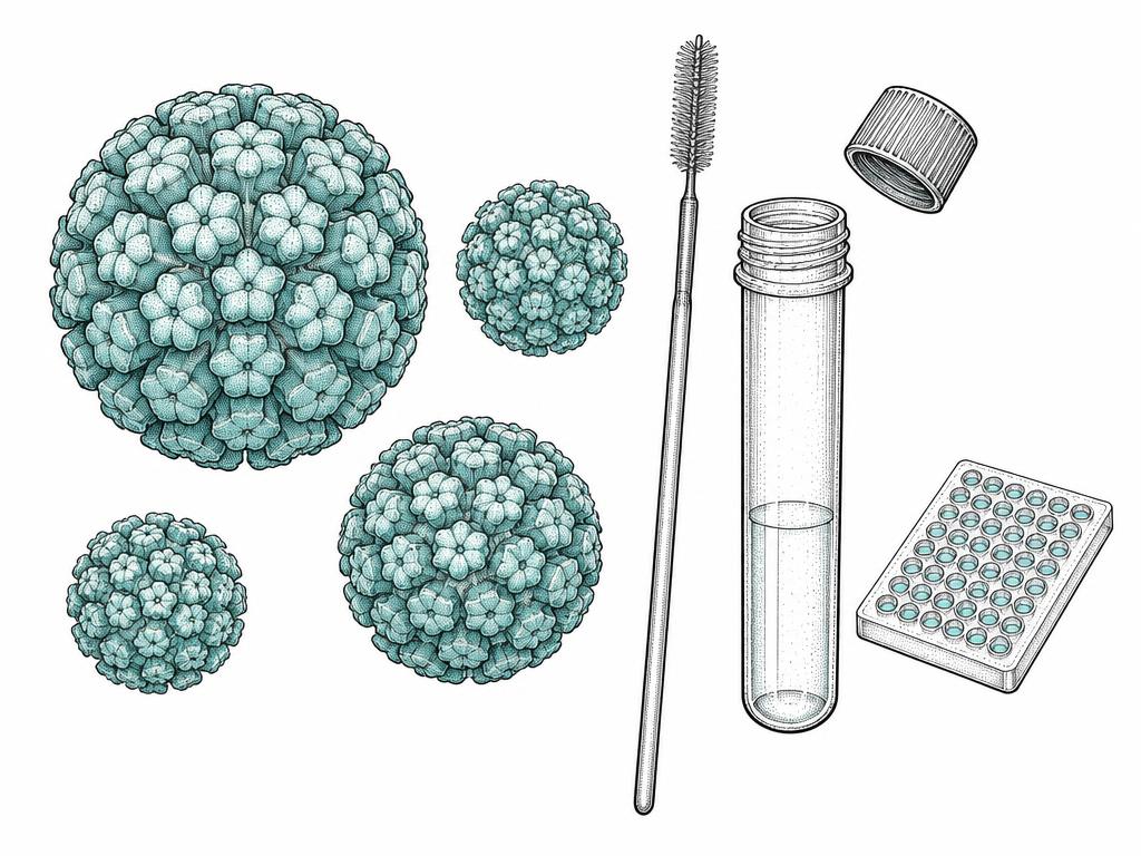
Human papillomaviruses (HPV) are non-enveloped viruses with a circular, double-stranded DNA genome. More than 230 types have been described, a little over 40 of which occur in the anogenital region. Based on their oncogenic potential they are divided into high-risk and low-risk types.
Sample collection
For testing, cell material is collected with a small brush or spatula. The collection is quickly done and usually described as well tolerated; the material reaches the laboratory in a carrier fluid.
Molecular detection and typing
In the laboratory the viral DNA is usually detected by PCR. At the same time it can be determined whether a high-risk or a low-risk type is present. This result influences further work-up and the follow-up intervals.
Relevance for head and neck
Besides the anogenital region, high-risk types can also affect the oral cavity and pharynx. A substantial share of squamous cell carcinomas of the mouth and throat is caused by an HPV infection. Abnormal findings in this area are therefore medically evaluated.
Frequently asked questions
This general information does not replace individual medical advice. Follow the instructions of your examining team.
Medical information and sources
12
Virus diagnostics
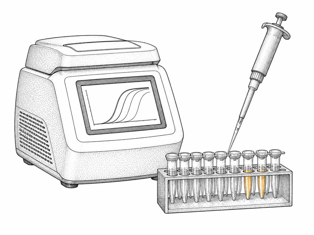
Direct detection of a pathogen is now largely achieved by molecular methods. They examine a sample for viral genetic material and are suitable for viruses that occur in the head and neck region – for example herpes simplex or varicella zoster viruses.
PCR as the method of first choice
Detecting viral DNA by PCR is regarded as the standard method for herpes simplex and varicella zoster viruses: it is particularly sensitive and specific, usually delivers results within 24 hours and distinguishes the virus types from one another.
Antibodies, culture and rapid tests
Antibody tests in the blood indicate a past or current encounter with the pathogen but are less precise for dating it. Virus culture and direct fluorescence labelling work mainly in early lesions.
When a test is useful
A test is especially helpful when therapy depends on it – for example in facial herpes zoster, in sudden hearing or balance changes with blisters, in suspected virus-related facial palsy, or in smell and taste disorders after infections. The decision is made by the examining physician based on findings and course.
Frequently asked questions
This general information does not replace individual medical advice. Follow the instructions of your examining team.
13
ECG (electrocardiogram)
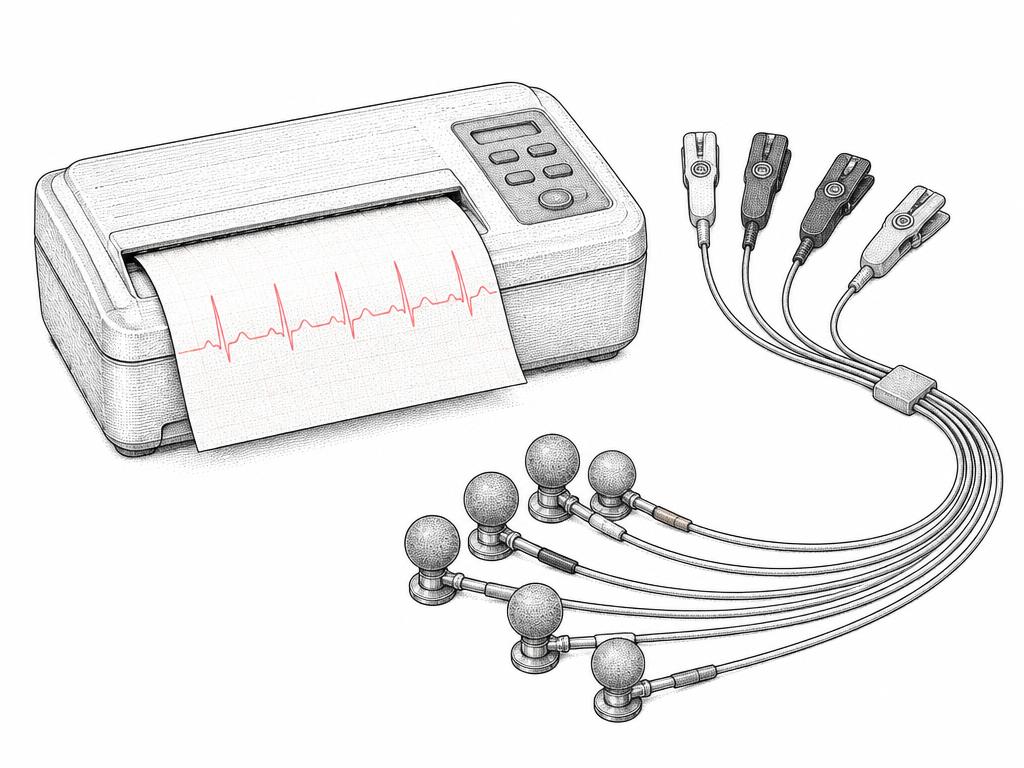
The ECG records the electrical activity of the heart. Electrodes on the skin pass the signals to a device that displays them as curves. It is usually the first and quickest examination when the heart’s capacity for strain needs to be assessed.
Procedure
For a resting ECG you lie relaxed on a couch. Small electrodes are attached to the chest, arms and legs and connected by cables to the ECG device; the recording takes only a few minutes.
What the curves show
The deflections of the curve follow the heart’s sequence of excitation. From their height, steepness, duration and time intervals, rhythm, rate and disturbances of impulse formation can be assessed.
Why the ECG matters here
Before an anaesthetic and an inpatient stay, the question of the heart’s capacity for strain is part of the work-up. In cases of racing heart, palpitations, dizziness or breathlessness the ECG is supplemented – where appropriate by a long-term or exercise ECG.
Frequently asked questions
This general information does not replace individual medical advice. Follow the instructions of your examining team.
Medical information and sources
14
Lung function and spirometry
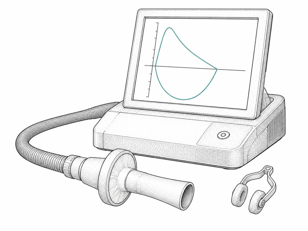
In spirometry you breathe through a mouthpiece into a measuring device while the nose is closed with a clip. The device measures the volumes of air you inhale and exhale and how fast the exhalation proceeds. The test is not painful but does require your active cooperation.
Procedure
After a short instruction you breathe in deeply and blow the air out into the mouthpiece as forcefully and as long as possible. The measurement is repeated several times; it usually takes less than ten minutes.
The key values
The main measures are the amount of air exhaled in the first second (FEV1) and the total amount exhaled (FVC). From their ratio, physicians can tell whether the airways are narrowed or the lung volume is reduced.
When a measurement is useful
Reasons include unexplained breathlessness, persistent cough or sputum, follow-up in asthma or COPD, and risk assessment before an operation. Within sleep medicine, lung function can also be assessed as a complement.
Frequently asked questions
This general information does not replace individual medical advice. Follow the instructions of your examining team.
After the examination
The results are discussed at the clinic. The next steps are agreed with the patient — from observation through conservative treatment to surgery.
Typical reasons
The treatment areas on this website describe the symptoms for which an assessment is advisable: sudden hearing changes and tinnitus, blocked nasal breathing, recurrent infections, snoring with breathing pauses and skin changes. When in doubt, an initial consultation clarifies whether further examination is required.