
Kopfzentrum Bielefeld
Cholesteatoma
Diagnosis
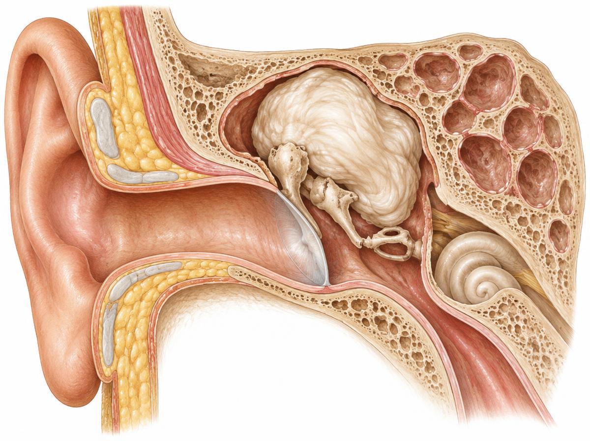
A cholesteatoma is a collection of keratinising skin cells in the middle ear and mastoid that grows slowly and can damage surrounding structures. The diagnosis is usually made under the operating microscope or with an endoscope: the eardrum is retracted in the attic region or shows a perforation through which a whitish mass is visible. A hearing test shows whether the ossicles are already involved; computed tomography can show the extent and bone involvement in more detail.
Symptoms
Typically there is a persistent, often foul-smelling discharge from one ear, frequently accompanied by progressive hearing loss or a feeling of pressure. Because the mass can attack bony structures, dizziness, noises in the ear or weakness of the facial muscles are warning signs that should be assessed promptly.
Causes
A cholesteatoma most often develops from a long-standing ventilation disorder of the eustachian tube: the eardrum retracts in the attic region and forms a pocket in which skin cells collect. After eardrum perforations or operations, skin can grow into the middle ear; a congenital form is observed rarely.
Treatment options
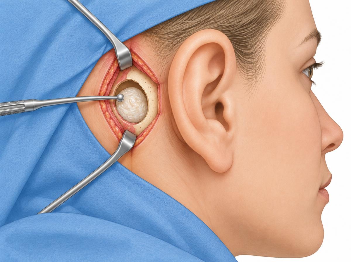
A cholesteatoma cannot be removed with drops or medication; treatment is surgical. During the operation the mass is removed through an approach behind the ear and the mastoid is cleared; procedures differ in whether the back wall of the ear canal is preserved or a permanently open cavity is created. Damaged ossicles can be reconstructed in the same or a later session with a tympanoplasty. Because regrowth is possible, follow-up appointments over many years are recommended.
After the procedure
After the operation the wound is cared for and protected for several weeks; the ear should not be submerged in water during this time. Hearing tests and microscopic examinations accompany healing. Sometimes a second operation follows after a few months to check the result, remove residual tissue or improve hearing.