
Kopfzentrum Bielefeld
Cholesterol granuloma of the petrous apex
Diagnosis
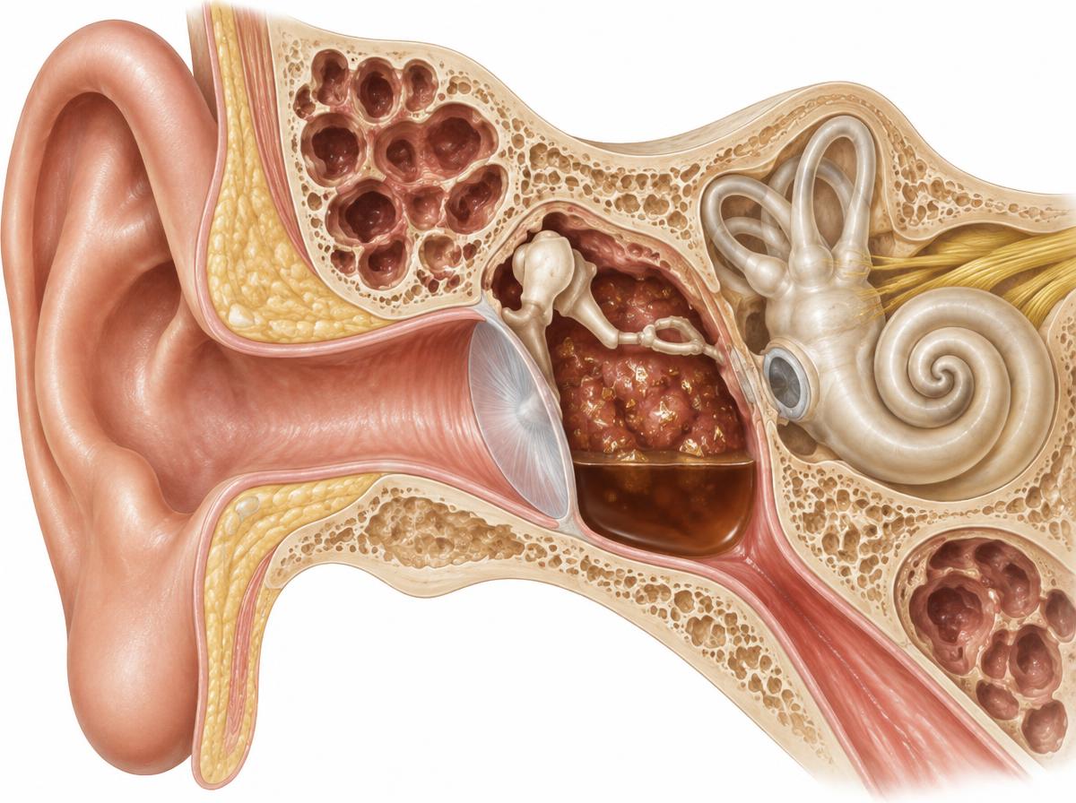
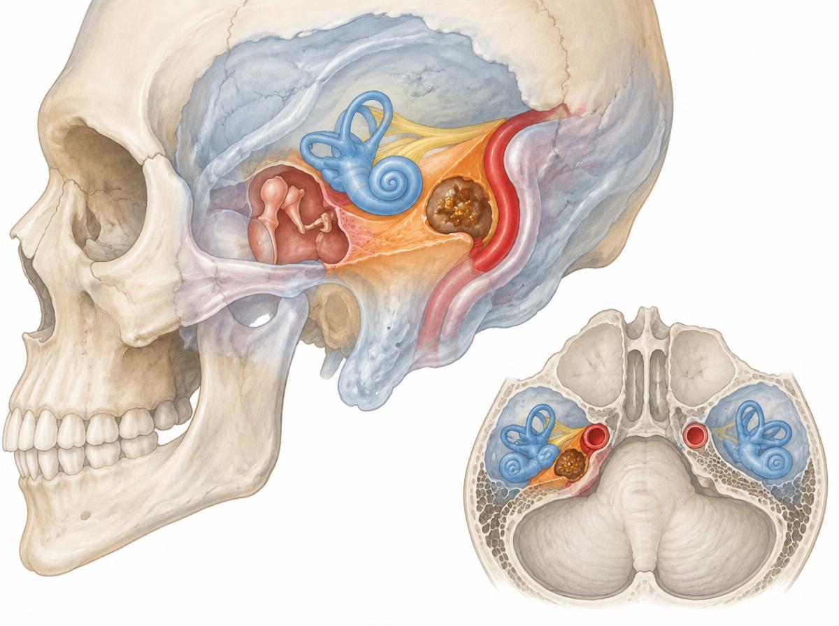
A cholesterol granuloma is a slowly expanding cyst filled with cholesterol crystals in the mastoid or at the petrous apex. Under the ear microscope it appears as a dark brown to bluish shimmering area behind an intact eardrum; a hearing test can show a conductive component. Magnetic resonance imaging provides the decisive picture: the finding is bright in both sequences, which distinguishes it from other mass lesions.
Symptoms
Many granulomas are discovered incidentally on imaging. When symptoms occur, they are mostly a feeling of pressure, slow hearing loss or noises in the ear; with deeply located findings at the petrous apex, double vision or facial numbness can occur when nerves are compressed.
Causes
The granuloma forms when air cells of the petrous bone are filled with blood or inflammatory fluid and are no longer ventilated: cells break down the blood, released cholesterol crystals irritate the tissue, and a cyst filled with crystals and inflammatory cells grows slowly.
Treatment options
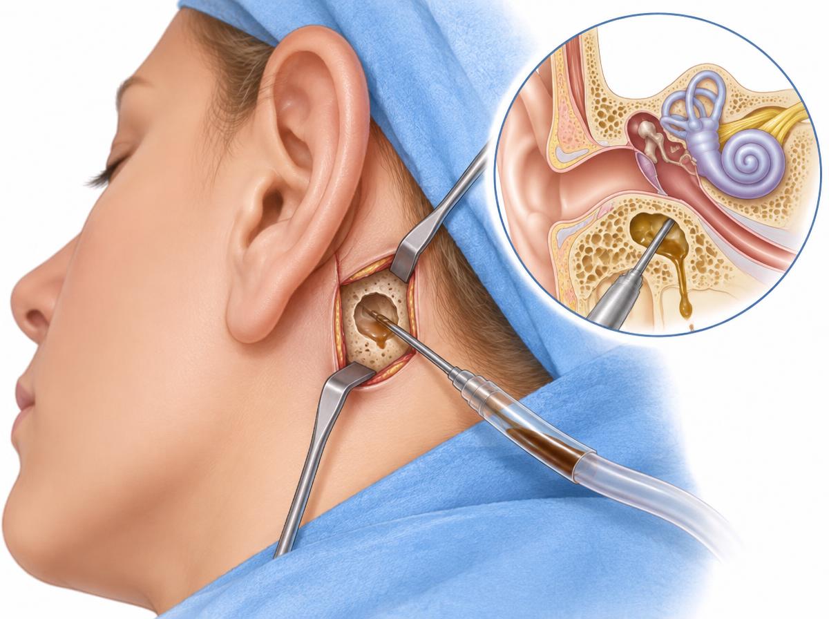
A small, unremarkable finding can be observed with imaging controls. Symptoms or growth indicate a draining operation: through a small bone window below the cochlea (infracochlear approach) the cyst is opened and permanently connected to the middle ear or the ear canal with a fine tube, so that the fluid does not collect again. The approach preserves hearing and the facial nerve and is reached from the ear canal.
After the procedure
The wound behind the ear usually heals within two weeks. The drainage tube remains in place permanently and is checked for patency in follow-up examinations; imaging shows that the cyst shrinks. Hearing tests document that hearing is preserved.