
Kopfzentrum Bielefeld
Cerebrospinal fluid leak with transnasal coverage
Diagnosis
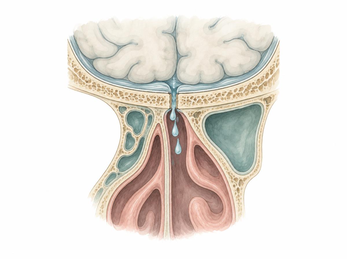
In a cerebrospinal fluid (CSF) leak, brain fluid escapes through an opening in the skull base and its meningeal covering – in the forms treated through the nose, into the nasal cavity or the paranasal sinuses. Suspicion usually arises from one-sided, clear, watery discharge from the nose. To confirm it, the fluid is tested for beta-2 transferrin. High-resolution computed tomography and magnetic resonance cisternography together show where the opening is and how large it is.
Symptoms
Typical is a one-sided, clear, watery running sensation from the nose that may worsen when lying down, bending forward or straining. Many people notice a salty or metallic taste, a running down the throat, or pressure in the head. Fever, neck stiffness and severe headache can indicate meningitis – in this case immediate medical attention is required.
Causes
Most often a CSF leak follows a head or facial injury; less often it follows a procedure on the paranasal sinuses or the skull base. Some leaks appear without any external trigger, for example with persistently raised intracranial pressure, congenital gaps in the skull base, or diseases that erode the bone of the skull base.
Treatment options
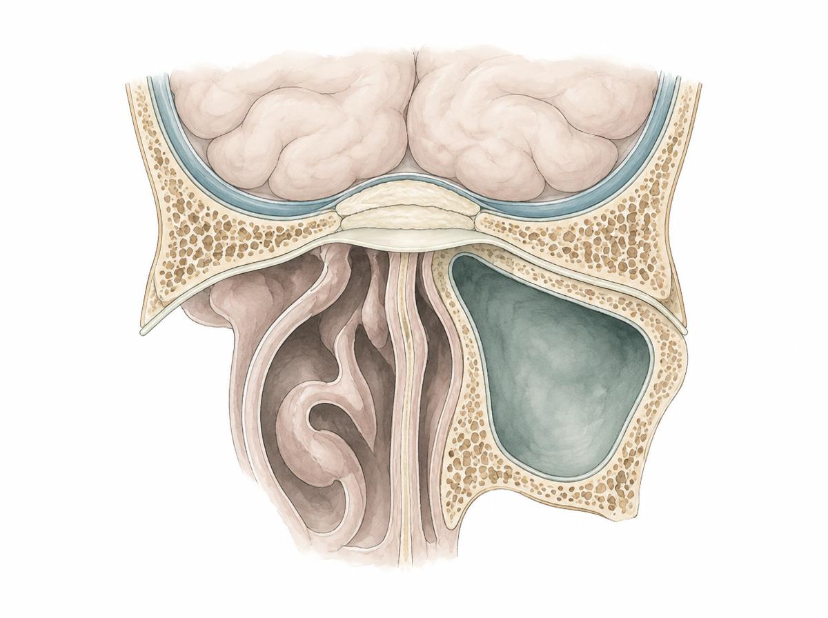
Small, recently formed leaks sometimes stop with rest and avoidance of straining. If CSF continues to flow, the opening is usually closed endoscopically through the nose: after gentle preparation of the surroundings, the site is covered from the nasal side in several layers, for example with a connective tissue layer, mucosa, fat, or a combination. In selected cases a temporary lumbar drainage of CSF supports the coverage until the seal is watertight. For very large or hard-to-reach defects, an open approach over the skull base may be required.
Endoscopic transnasal closure is regarded as highly reliable: in a review of 289 CSF fistulae repaired in this way, the first procedure succeeded in about nine out of ten cases, and overall about 97 percent of the leaks could be closed by an endoscopic approach. After the operation come follow-up examinations and a period without straining, lifting and flying. If the coverage does not seal permanently, it can be placed again.