
Kopfzentrum Bielefeld
Median and lateral neck fistulas
Diagnosis
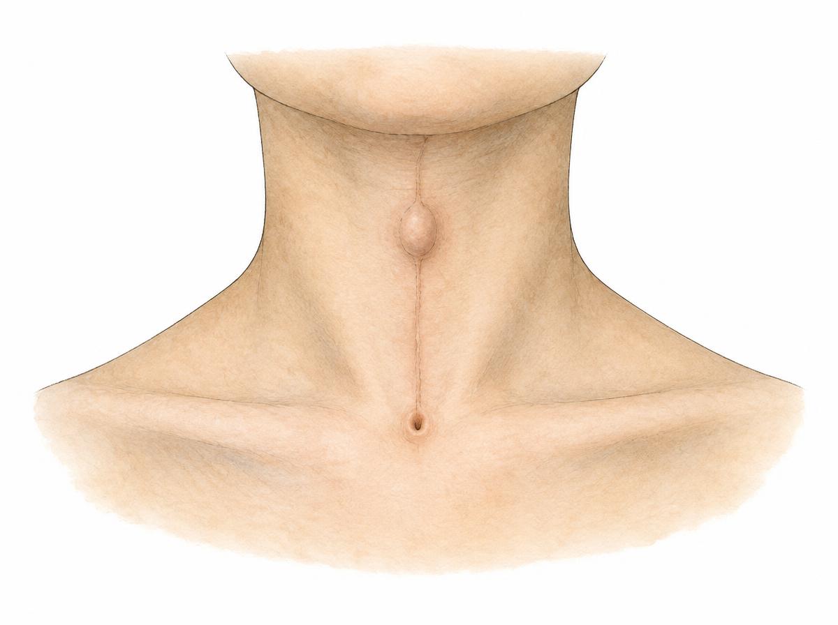
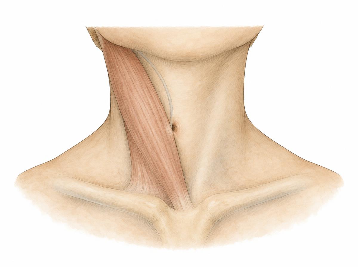
Median and lateral neck fistulas are congenital tracts left behind by the development of the thyroid gland and the neck. The median neck fistula follows the path the thyroid took from the base of the tongue into the lower neck and lies in the midline; the lateral neck fistula follows the remnants of the branchial apparatus and lies at the front edge of the large neck muscle. The skin opening and its secretion are assessed; an ultrasound study, more rarely magnetic resonance imaging, shows the course and extent. With a median finding it is also checked whether functioning thyroid tissue is present.
Symptoms
Median neck fistulas appear as a small opening or as a firm, mobile swelling in the midline of the neck, usually just above the voice box; the swelling moves with swallowing or with sticking out the tongue. Lateral neck fistulas appear as a small opening at the front edge of the large neck muscle, from which clear or milky secretion escapes. Both forms can become inflamed, swell and form pus; after a healed collection, a palpable cord or a scar often remains.
Causes
Both arise during embryonic development: the tract the thyroid takes on its way from the base of the tongue into the lower neck does not close completely. If parts of the branchial arches, from which the neck and the ears develop, do not regress, they remain as a tract or a cyst. Median findings usually appear in childhood before the age of ten, lateral ones more often in children and young adults.
Treatment options
If the tract is inflamed or purulent, the inflammation is treated first, if necessary by opening a collection and with antibiotics, until the finding settles. Median neck fistulas are removed surgically, with part of the hyoid bone and the tract up to the base of the tongue taken along, so that the finding does not reform. Lateral neck fistulas are removed as a complete tract with the skin opening and the inner continuation. Follow-up examinations come after the procedure; if tissue remains, a further procedure may be required.